AbstractObjectivesThe purpose of this study is to examine the effect of water and fermentation extracts of KMS (Kami-Mihudeongsikjang-tang) on AD (atopic dermatitis). Additionally, by applying the fermentation extracts of KMS at the first sensitization and latency period, I investgated whether it could prevent AD.
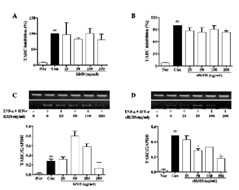
MethodsIn this study, to test the effect and preventive efficacy of KMSs on AD. DNCB-induced BALB/c mice of AD model was used. Through histological observation, epidermis and dermis thickness, the infiltration of eoshiphils and mast cells in epidermis and dermis were examined. We measured the serum level of IgE and IL-6, TNF-alpha, and the expressions of NF-κB and MAPK protein. In order to examine the effect of KMSs on keratinocyte, HaCaT cells were treated TNF-alpha and IFN-gamma to induce an inflammatory response. The KMSs were applied at various concentration in the experimental group. We investigated TARC expression.
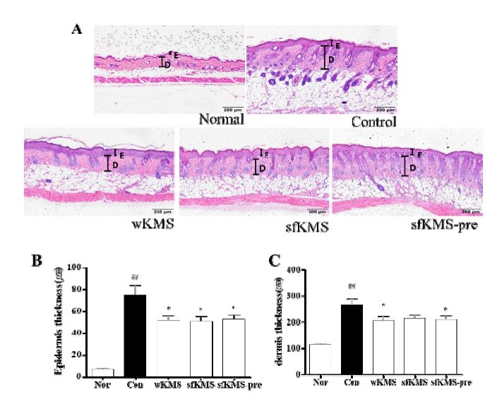
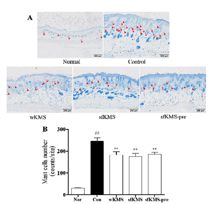
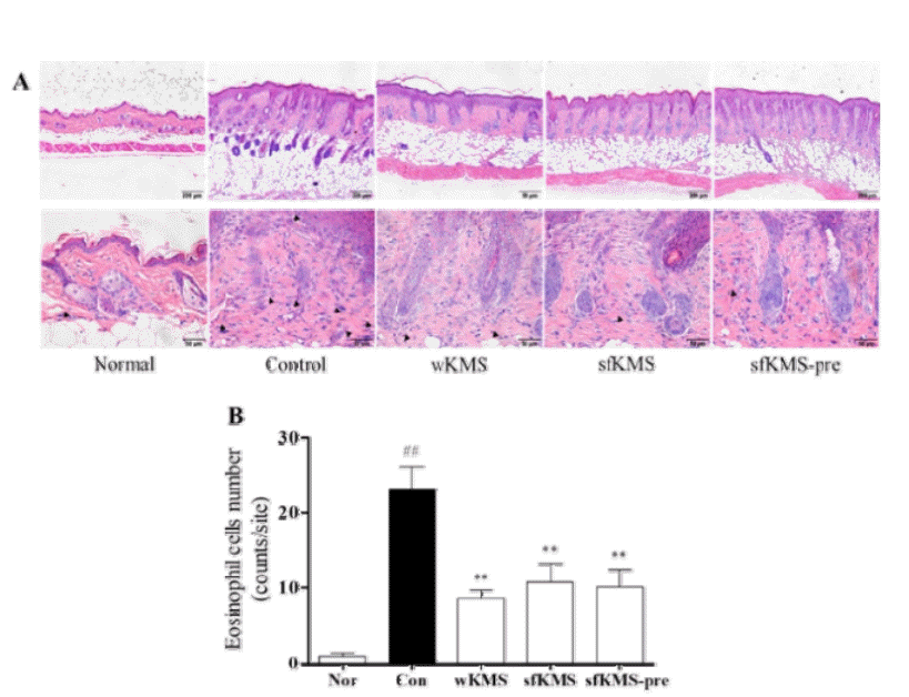
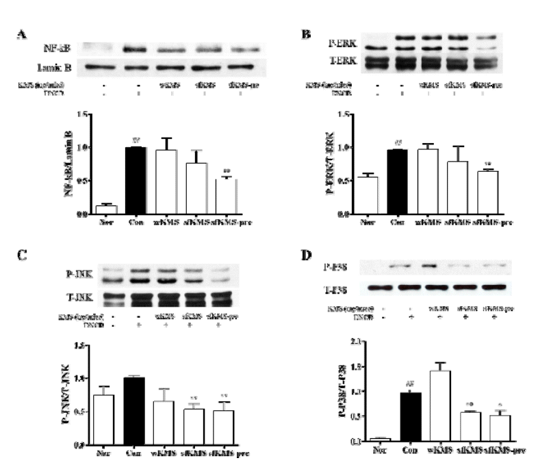
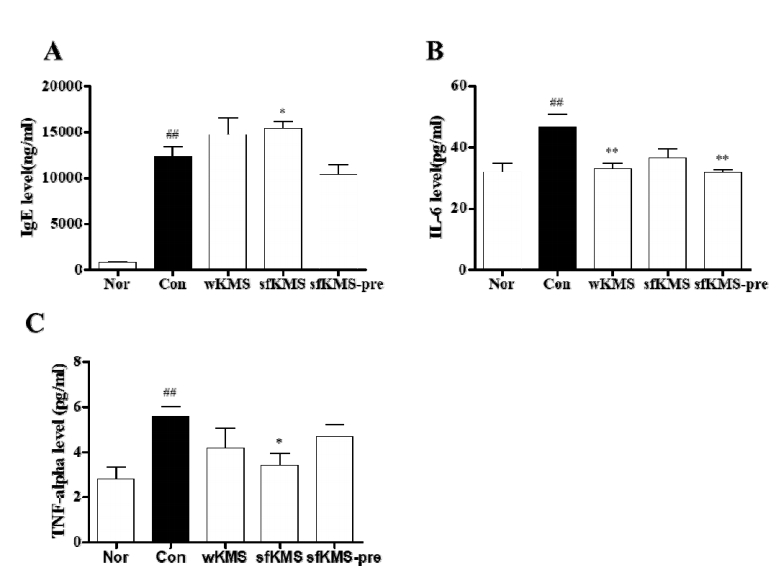
ResultsThe treatment groups were reduced epidermis and dermis thickness, inhibited the infiltration of eosinophils and mast cells, reduced the serum level of IL-6. Moreover, sfKMS group reduced serum level of TNF-alpha, inhibited the protein expressions of NF-κB and the phosphoryllation of ERK, JNK and p38. Especially sfKMS-pre group were reduced the serum level of IgE, show significant inhibition on the protein expression of NF-κB and the phosphoryllation of ERK, JNK and p38. In the experiment on HaCaT cells, sfKMS group were reduced expression of TARC.
참고문헌1. Coca AF, Cooke RA. On the classification of the phenomena of hypersensitiveness. J Immunol. 1923; 8:163–182.
2. Nutten S. Atopic dermatitis: global epidemiology and risk factors. Ann Nutr Metab. 2015; 66:1. 8–16.
3. Lee YU, Park SH, Nam EH, Chung TH, Hwang CY. Modulatory effects of PG102 (Actinidia arguta) on inflammatory cytokine mRNA expression in canine atopic dermatitis. Thai J Vet Med. 2016; 46:4. 601–607.
4. Marco M, Heeney D, Binda S, Cifelli CJ, Cotter PD, Foligne B, et al. Health benefits of fermented foods: microbiota and beyond. Current Opinion in Biotechnology. 2017; 44:94–102.
5. Um YR, Lee JH, Ma JY. Quantitative analysis of marker substances in Solid fermented Angelicae Gigantis Radis by HPLC. Korean Journal of Oriental Medicine. 2010; 16:1. 173–178.
6. Wickman M, Asarnoj A, Tillander H, Andersson N, Bergstrom A, Kull I, et al. Childhood-to -adolescence evolution of IgE antibodies to pollens and plant foods in the BAMSE cohort. J Allergy Clin Immunol. 2014; 133:2. 580–582.
7. Westman M, Asarnoj A, Hamsten C, Wickman M, van Hage M. Windows of opportunity for tolerance induction for allergy by studying the evolution of allergic sensitization in birth cohorts. Seminars in Immunology. 2017; 30:61–66.
8. Peng W, Novak N. Pathogenesis of atopic dermatitis. Clinical & Experimental Allergy. 2015; 45:566–574.
9. Bieber T. Atopic Dermatitis. Ann Dermatol. 2010; 22:125–137.
10. Kasutani K, Fujii E, Ohyama S, Adachi G, Hasegawa M, Kitamura H, Yamashita N. Anti-IL-31 receptor antibody is shown to be a potential therapeutic option for treating itch and dermatitis in mice. Br J Pharmacol. 2014; 171:5049–5058.
11. Mu Z, Zhao Y, Liu X, Chang C, Zhang J. Molecular biology of atopic dermatitis. Clin Rev Allergy Immunol. 2014; 47:193–218.
12. Johansson SG, Bieber T, Dahl R, Friedmann PS, Lanier BQ, et al. Revised nomenclature for allergy for global use: Report of the Nomenclature Review Committee of the World Allergy Organization. J Allergy Clin Immunol. 2004; 113:832–836.
13. Ong PY, Leung DY. Immune dysregulation in atopic dermatitis. Curr Allergy Asthma Rep. 2006; 6:384–389.
14. Leung DY, Boguniewicz M, Howell M, Nomura I, Hamid QA. New insights into atopic dermatitis. J Clin Invest. 2004; 113:651–657.
15. Mosmann TR, Cherwinski H, Bond MW, Giedlin MA, Coffman RL. Two types of murine helper T cell clone. I. Definition according to profiles of lymphokine activities and secreted proteins. J Immunol. 1986; 136:7. 2348–2357.
16. Maggi E, Prete GD, Macchia D, Parronchi P, Tiri A, Chrétien I, et al. Profiles of lymphokine activities and helper function for IgE in human T cell clones. Eur J Immunol. 1988; 18:1045–1052.
17. Robinson DS, Hamid Q, Ying S, Tsicopoulos A, Barkans J, Bentley AM, et al. Predominant TH2-like bronchoalveolar Tlymphocyte population in atopic asthma. N Engl J Med. 1992; 326:298–304.
18. Chan LS, Robinson N, Xu L. Expression of interleukin-4 in the epidermis of transgenic mice results in a pruritic inflammatory skin disease: an experimental animal model to study atopic dermatitis. J Invest Dermatol. 2001; 117:977–983.
19. Pulendran B, Artis D. New paradigms in type 2 immunity. Science. 2012; 337:431–435.
20. Hirano T. Interleukin 6 and its receptor: 10 years later. Int Rev Immunol. 1998; 16:249–284.
21. Diehl S, Rincón M. The two faces of IL-6 on Th1/Th2 differentiation. Mol Immunol. 2002; 39:9. 531–36.
22. Danso MO, van Drongelen V, Mulder A, van Esch J, Scott H, van Smeden J, et al. TNF-a and Th2 Cytokines Induce Atopic Dermatitis - Like Features on Epidermal Differentiation Proteins and Stratum Corneum Lipids in Human Skin Equivalents. Journal of Investigative Dermatology. 2014; 134:1941–1950.
23. Cameron DJ, Tong Z, Yang Z, Kaminoh J, Kamiyah S, Chen H, et al. Essential role of Elovl4 in very long chain fatty acid synthesis, skin permeability barrier function, and neonatal survival. Int J Biol Sci. 2007; 3:111–119.
24. van Smeden J, Janssens M, Kaye EC, Caspers PJ, Lavrijsen AP. The importance of free fatty acid chain length for the skin barrier function in atopic eczema patients. Exp Dermatol. 2014; 23:45–52.
25. Zhu Z, Oh M-H, Yu J, Liu YJ, Zheng T. The Role of TSLP in IL-13-induced atopic march. Sci Rep. 2011; 1:23
26. Karin M, Ben-Neriah Y. Phosphorylation meets ubiquitination: The control of NF-kB activity. Annu Rev Immunol. 2000; 18:621–663.
27. Aupperle K, Bennett B, Han Z, Boyle D, Manning A, Firestein G. NF-k B regulation by I k B kinase-2 in rheumatoid arthritis synoviocytes. J Immunol. 2001; 166:2705–2711.
28. Monaco C, Andreakos E, Kiriakidis S, Mauri C, Bicknell C, Foxwell B, et al. Canonical pathway of nuclear factor k B activation selectively regulates proinflammatory and prothrombotic responses in human atherosclerosis. Proc Natl Acad Sci U S A. 2004; 101:5634–5639.
29. Zhang YL, Dong C. MAPkinase sinimmuneresponses. Cell Mol Immunol. 2005; 2:20–27.
30. Vestergaard C, Yoneyama H, Murai M, Nakamura K, Tamaki K, Terashima Y, et al. Overexpression of Th2-specific chemokines in NC/Nga mice exhibiting atopic dermatitis-like lesions. J Clin Invest. 1999; 104:1097–1105.
31. Sallusto F, Lenig D, Mackay CR, Lanzaveccchia A. Flexible programs of chemokine receptors expression on human polarized T helper 1 and 2 lymphocytes. J Exp Med. 1998; 187:875–883.
32. Vestergaard C, Bang K, Gesser B, Yoneyama H, Matsushima K, Larsen CG. A Th2 chemokine, TARC, prouduced by keratinocytes may recruit CLA+CCR4+ lymphocytes into lesional atopic dermatitis skin. J Invest Dermatiol. 2000; 115:640–646.
33. Kakinuma T, Nakamura K, Wakugawa M, Mitsui H, Tada Y, Saeki H, et al. Thymus and activation-regulated chemokine in atopic dermatitis: serum thymus and activation-regulated chemokine level is closely related with disease activity. J Allergy Clin Immunol. 2001; 107:535–541.
|
|
|||||||||||||||||||||||||||||||
|
|||||