Efficacy of Electroacupuncture with Dysphagia Caused by Stroke:

Double-blind, Randomized Controlled Trial

Article information

J Korean Med. 2014;35(1):26-40
Publication date (electronic) : 2014 March 30
doi : https://doi.org/10.13048/jkm.14003
Department of Cardiology and Neurology, College of Korean Medicine, Kyung Hee University
Correspondence to:박성욱(Seong-Uk Park), 서울시 강동구 동남로 892 강동경희대학교 병원 중풍뇌질환센터 한방내과, Tel: +82-2-440-6217, Fax: +82-2-440-6217, E-mail: seonguk.kr@gmail.com
Received 2013 September 11; Revised 2013 December 30; Accepted 2013 December 30.

Abstract

Objectives

This study investigated the effect of electroacupuncture on swallow function in stroke patients with dysphagia. The purpose was to determine whether electroacupuncture could improve swallow function and quality of life for patients with dysphagia caused by stroke. A pilot double-blind, randomized controlled trial design was used

Methods:

A total of 17 stroke patients with dysphagia were recruited to this study, 8 assigned to the swallowing electroacupuncture (SE) group and 9 to the control group. Swallowing electroacupuncture was assessed for 5 Hz electrical or sham stimulation for 20 min duration 3 times a week. Outcome measurements were DOSS, 14-item questionnaire from the SWAL-QOL, NIHSS and MBI. Assessment was carried out for baseline, 2 weeks and 4 weeks

Results:

In the 2 weeks follow up data, the study group showed significant difference in DOSS and SWAL-QOL compared with the baseline. The study group also showed significant difference in DOSS and SWAL-QOL compared with the control group. Similarly in the 4 weeks follow up data, the study group showed improvement tendency in DOSS and SWAL-QOL compared with baseline and also with the control. The NIHSS and MBI scores returned no significant differences. The swallowing electroacupuncture was well tolerated in all cases with no serious adverse effects.

Conclusions:

The findings from the pilot study indicated that electroacupuncture has significant effects on improvement in swallowing function and quality of life. With both inpatients and outpatients, further larger and longer-term follow-up study is needed to confirm this suggestion.

Fig. 1

Randomization and Patients Flow Chart

Baseline Characteristics of Patients

Changes in Dysphagia Outcome and Severity Scale

Changes in Swallowing Quality of Life

Changes in National Institutes of Health Stroke Scale

Changes in Modified Barthel Index

References

1. Marik PE, Kaplan D. Aspiration pneumonia and dysphagia in the elderly. Chest 2003;124:328–36.
2. Mann G, Hankey GJ, Cameron D. Swallowing Function After Stroke Prognosis and Prognostic Factors at 6 Months. Stroke 1999;30:744–8.
3. Sharma JC, Fletcher S, Vassallo M, Ross I. What influences outcome of stroke-pyrexia or dysphagia? Int J Clin Pract 2001;55:17–20.
4. Martino R, Foley N, Bhogal S, Diamant N, Speechley M, Teasell R. Dysphagia After Stroke Incidence, Diagnosis, and Pulmonary Complications. Stroke 2005;36:2756–63.
5. Dziewas R, Ritter M, Schilling M, Konrad C, Oelenberg S, Nabavi DG, et al. Pneumonia in acute stroke patients fed by nasogastric tube. J Neurol Neurosurg Psychiatr 2004;75:852–6.
6. Langmore SE, Terpenning MS, Schork A, Chen Y, Murray JT, Lopatin D, et al. Predictors of aspiration pneumonia: how important is dysphagia? Dysphagia 1998;13:69–81.
7. Lim KB, Lee HJ, Lim SS, Choi YI. Neuromuscular electrical and thermal-tactile stimulation for dysphagia caused by stroke: a randomized controlled trial. Journal of Rehabilitation Medicie 2009;41:17–48.
8. Long YB, Wu XP. A meta-analysis of the efficacy of acupuncture in treating dysphagia in patients with a stroke. Acupunct Med 2012;30(4):291–7.
9. Gomez-Busto F, Andia Munoz V, Sarabia M, Ruiz de Alegria L, González de Viñaspre I, López-Molina N, et al. Gelatinous nutritional supplements: a useful alternative in dysphagia. Nutr Hosp 2011;26:775–83.
10. Seki T, Iwasaki K, Arai H, Sasaki H, Hayashi H, Yamada S, et al. Acupuncture for dysphagia in poststroke patients: a videofluoroscopic study. J Am Geriatr Soc 2005;53:108–34.
11. Seki T, Kurusu M, Tanji H, Arai H, Sasaki H. Acupuncture and swallowing reflex in poststroke patients. J Am Geriatr Soc 2003;51:72–67.
12. Kim TH, Na BJ, Rhee JW, Lee CR, Park YM, Choi CM, et al. The Effect of Moxibustion at Chonjung(CV17, Shanzhong) on Patients with Dysphagia after Stroke. 2005;
13. Iwasaki K, Kato S, Monma Y, Niu K, Ohrui T, Okitsu R, et al. A Pilot Study of Banxia Houpu Tang, a Traditional Chinese Medicine, for Reducing Pneumonia Risk in Older Adults with Dementia. Journal of the American Geriatrics Society 2007;55:2035–40.
14. Ertekin C, Aydogdu I, Yüceyar N, Kiylioglu N, Tarlaci S, Uludag B. Pathophysiological mechanisms of oropharyngeal dysphagia in amyotrophic lateral sclerosis. Brain 2000;123:125–40.
15. Kang YW, Na DL, Hahn SH. A validity study on the Korean Mini-Mental State Examination (K-MMSE) in Dementia Patients. J Korean Neurol Assoc 1997;15(2):300–8.
16. O’Neil KH, Purdy M, Falk J, Gallo L. The dysphagia outcome and severity scale. Dysphagia 1999;14:139–45.
17. McHorney CA, Robbins J, Lomax K, Rosenbek JC, Chignell K, Kramer AE, et al. The SWAL-QOL and SWAL-CARE Outcomes Tool for Oropharyngeal Dysphagia in Adults: III. Documentation of Reliability and Validity. Dysphagia 2002;17:97–114.
18. McHorney CA, Bricker DE, Kramer AE, Rosenbek JC, Robbins J, Chignell KA, et al. The SWAL-QOL outcomes tool for oropharyngeal dysphagia in adults: I. Conceptual foundation and item development. Dysphagia 2000;15:115–21.
19. McHorney CA, Bricker DE, Robbins J, Kramer AE, Rosenbek JC, Chignell KA. The SWAL-QOL outcomes tool for oropharyngeal dysphagia in adults: II. Item reduction and preliminary scaling. Dysphagia 2000;15:122–33.
20. Kasner SE. Clinical interpretation and use of stroke scales. The Lancet Neurology 2006;5:603–12.
21. Hocking C1, Williams M, Broad J, Baskett J. Sensitivity of Shah, Vanclay and Cooper’s modified Barthel Index. Clin Rehabil 1999;13(2):141–7.
22. Terre R, Mearin F. Oropharyngeal dysphagia after the acute phase of stroke: predictors of aspiration. Neurogastroenterol Motil 2006;18:200–5.
23. Barer DH. The natural history and functional consequences of dysphagia after hemispheric stroke. J Neurol Neurosurg Psychiatr 1989;52:236–41.
24. Smithard DG, O’Neill PA, England RE, Park CL, Wyatt R, Martin DF, et al. The natural history of dysphagia following a stroke. Dysphagia 1997;12:188–93.
25. Park CL, O’Neill PA, Martin DF. A pilot exploratory study of oral electrical stimulation on swallow function following stroke: an innovative technique. Dysphagia 1997;12:161–6.
26. Carnaby-Mann GDCM. Examining the evidence on neuromuscular electrical stimulation for swallowing: A meta-analysis. 2007;133:564–71.
27. Fraser C, Power M, Hamdy S, Rothwell J, Hobday D, Hollander I, et al. Driving Plasticity in Human Adult Motor Cortex Is Associated with Improved Motor Function after Brain Injury. Neuron 2002;34:831–40.
28. Li KL. Treatment of limb paralysis using low-frequency deep electric stimulation. Med Tr Prom Ekol 1995;(9):33–7.
Article information Continued

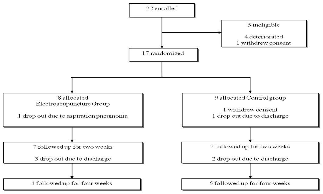
Fig. 1

Randomization and Patients Flow Chart

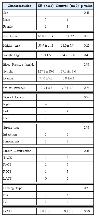
Table 1

Baseline Characteristics of Patients

Characteristics SE (n=8) Control (n=9) p-value
Sex 0.48
  Male 7 6
  Female 1 3

Age (years) 65.8±11.4 70.7±9.2 0.32

Height (cm) 58.9±11.8 68.0±9.8 0.22

Weight (kg) 170.3±5.3 164.7±7.6 0.40

Blood Pressure (mmHg) 0.88
  Systolic 127.8±20.6 127.1±18.6
  Diastolic 71.0±7.2 73.8±6.2

On set (weeks) 10.3±8.8 7.7±3.2 0.74

Side of Lesion 0.74
  Right 4 3
  Left 2 4
  Both 2 2

Stroke type 0.88
  Infarction 5 6
  Hemorrhage 3 3

Stroke Classification 0.48
  TACS 3 2
  PACS 2 2
  POCS 3 5
  LACS 0 0

Feeding Type 0.27
  NG 7 5
  PO 1 4

DOSS 2.8±1.4 3.0±1.3 0.70

Values are mean±SD, n

SE:Swallowing Electroacupuncture therapy group, TACS:total anterior circulation stroke, PACS:partial anterior circulation stroke, POCS:posterior circulation stroke, NG:Nasogastric tube feeding, PO: modified oral feeding, DOSS:dysphagia outcome and severity scale

Table 2

Changes in Dysphagia Outcome and Severity Scale

Baseline 2 weeks follow up 4 weeks follow up
Study group 2.8±1.4 3.9±0.9 (n=7) * 5.0±0.8 (n=4)
Control group 3.0±1.3 3.4±1.3 (n=7) 3.6±1.3 (n=5)

Values are mean±standard deviation

*

Indicates significant changes compared with baseline by Wilcoxon signed ranks test (p=0.03)

Indicates significant differences compared with control group by Mann-Whitney U test (p=0.04)

Table 3

Changes in Swallowing Quality of Life

Baseline 2 weeks follow up 4 weeks follow up
Study group 42.6±13.7 48.4±10.0 (n=7)* 53.8±9.5 (n=4)
Control group 47.5±12.7 48.0±9.4 (n=7) 55.4±5.8 (n=5)

Values are mean±standard deviation

*

Indicates significant changes compared with baseline by Wilcoxon signed ranks test (p=0.04)

Table 4

Changes in National Institutes of Health Stroke Scale

Baseline 2 weeks follow up 4 weeks follow up
Study group 11.3±4.1 10.9±3.4 (n=7) 11.0±2.0 (n=4)
Control group 11.2±4.4 9.0±3.0 (n=7) 8.6±2.9 (n=5)

Values are mean±standard deviation

Table 5

Changes in Modified Barthel Index

Baseline 2 weeks follow up 4 weeks follow up
Study group 5.3±6.6 7.1±6.4 (n=7) 5.5±3.0 (n=4)
Control group 4.1±4.5 4.4±4.9 (n=7) 5.0±5.7 (n=5)

Values are mean±standard deviation