Three Cases of Oral Mucoceles Treated with Traditional Korean Medicine
Article information
Abstract
This is the first case report about oral mucoceles treated with traditional Korean medication in Korea. One case of Blandin-Nuhn mucocele and two cases of ranula were treated with Ondam-tang and Dohongsamul-tang respectively. No recurrence of ranula was found for 30 months in one case and 3 months in other recent case. According to this result, we suppose that consideration of “Blood stasis and heat” might be helpful to treat progressed or large mucoceles while common mucoceles are generally regarded as results of “Phlegm heat”. We suggest that traditional Korean medication can be an alternative to preserve salivary glands and to minimize complications of surgery. Further studies with more cases and longer observation period should be performed to establish proper prescriptions for oral mucocele and the evidence of treatment rate and recurrence rate.
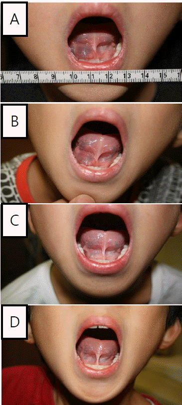
Photographs of Clinical Changes of Case 1.
A: 2013-05-11. Ranula on the right sublingual area, sized approximately 1.2cm×1.2cm.
B: 2013-08-31. No shrinkage was observed compared with A.
C: 2013-10-05. More than 50% shrinkage was observed.
D: 2013-11-26. The ranula was completely shrunk.
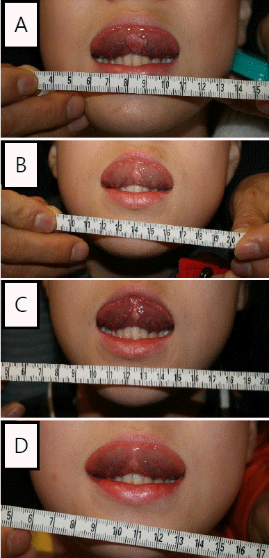
Photographs of Clinical Changes of Case 2.
A: 2015-11-23. Sublingual mucocele sized 1.0×1.1cm was observed.
B: 2015-12-12. The size was reduced to nearly 0.5×0.8cm.
C: 2016-01-02. The size was approximately 0.4×0.6cm.
D: 2016-01-23. The sublingual mucocele nearly disappeared.
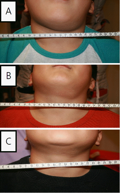
Photographs of Clinical Changes of Case 3.
A: 2016-01-23. Plunging ranula about 3cm diameter was observed in the left submandibular area.
B: 2016-02-04. The diameter of the plunging ranula decreased to about 2.0cm.
C: 2016-02-22. The mass was not observed.
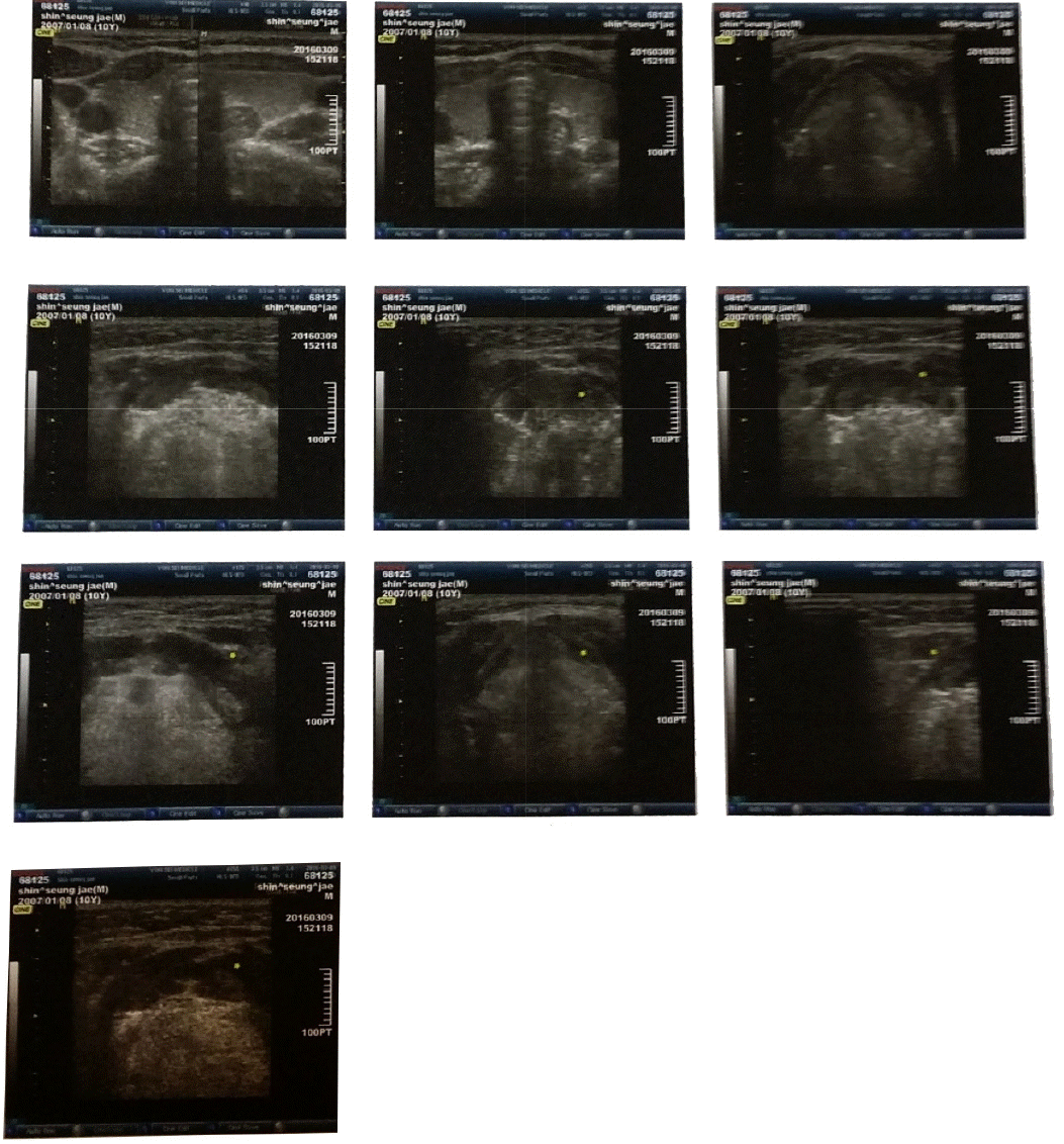
Ultrasonography of Case 3 on 2016-03-09.
The ultrasonography revealed there was no mass around the left submandibular gland. The picture only showed trace of inflammation with low-echo.