Protective effect of furosin isolated from Euphorbia helioscopia against glutamate-induced HT22 cell death

Article information

J Korean Med. 2018;39(1):35-43
Publication date (electronic) : 2018 March 31
doi : https://doi.org/10.13048/jkm.18004
1Kyeongnam National University of Science and Technology
2College of Korean Medicine, Gachon University
3Department of Oriental Neuropsychiatry, College of Korean Medicine, Gachon University
Correspondence to: 최성열 (Sung Youl Choi), 가천대학교 한의과대학 한방신경정신과, 1342 Seongnam-Daero, Sujung-Gu, Seongnam 13120, South Korea Tel: 82-32-770-1343, e-mail: pc1075@gachon.ac.kr
Received 2017 February 13; Revised 2018 March 13; Accepted 2018 March 14.

Abstract

Objectives

In the brain, glutamate is the most important excitable neurotransmitter in physiological and pathological conditions. However, the high level of glutamate induces neuronal cell death due to exitotoxicity and oxidative stress. The present study investigated to evaluate a possible neuroprotective effect of furosin isolated from Euphorbia helioscopia against glutamate-induced HT22 cell death.

Methods

Furosin was isolated from methanol extract of Euphorbia helioscopia and examined whether it protects glutamate-induced neuronal cell death. The cell viability was determined using Ez-Cytox assay. Anti-oxidative effect of furosin was determined by DPPH scavenging activities, and the levels of intracellular reactive oxygen species (ROS) were determined by the fluorescent intensity after staining the cells with H2DCFDA. To evaluate apoptotic cell death, we performed nuclear staining and image-based cytometeric analysis.

Results

The cell viability was significantly increased by treatement with furosin compared with the treatment with glutamate. Furosin showed a strong DPPH radical scavenging activity (EC50=1.83 μM) and prevented the accumulation of intra cellular ROS. Finally, the presence of 50 and 100 μM furosin significantly the percentage of apoptotic cells compared with glutamate treatment.

Conclusion

The present study found that furosin is a potent neuroprotectant against glutamate-induced oxidative stress through inhibition of apoptotic cell death induced by glutamate. Therefore, the present study suggests that furosin as a bioactive compound of E. helioscopia can be a useful source to develop a drug for the treatment of neurodegenerative diseases and acute brain injuries.
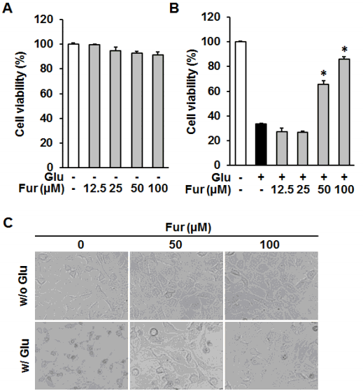
Fig. 1

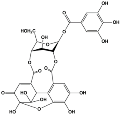
Chemical structure of furosin (Fur).

Fig. 2

Furosin inhibited glutamate-induced HT22 cell death. HT22 cells were treated with the indicated concentrations of furosin in the presence or absence of 5 mM glutamate. (A and B) To determine the self toxicity and protective effect of furosin, the cell viability was determined using Ez-Cytox assay kit after the exposure to glutamate for 24 h. Bars denote the percentage of cell viability compared with non-treated groups (mean±S.E.M., *p<0.05 compared with glutamate-treated group). (C)Morphological changes were observed under the phase contrast microscope.
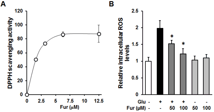
Fig. 3

Furosin prevented intracellular ROS accumulation through their antioxidant capacity. (A)The indicated concentrations of furosin was incubated with DPPH radical reagent for 30 min and obtained using microplate reader at 550 nm. (B)HT22 cells were treated with 50 or 100 uM furosin in the presence (dark gray) or absence (gray) of 5 mM glutamate for 8 h and stained with H2DCFDA, a fluorescent indicator of ROS. The fluorescent intensity of DCF was measured using a fluorescent microplate reader. Bars denote the fold increases of intracellular ROS level compared with non-treated groups (mean ±S.E.M., *<0.05 compared with glutamate-treated group).
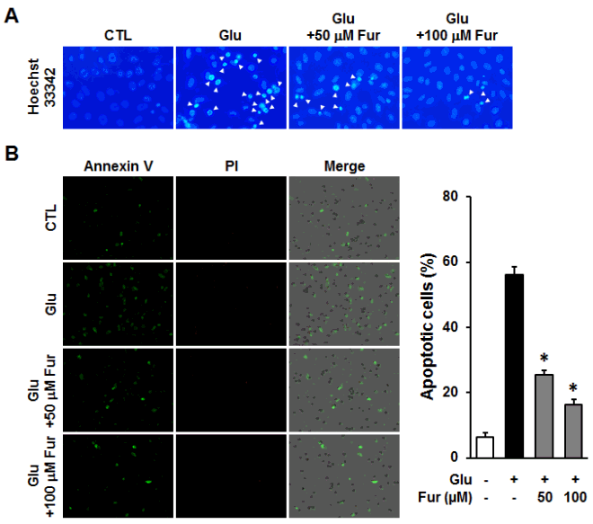
Fig. 4

Furosin prevented glutamate-induced apoptotic HT22 cell death. HT22 cells were incubated with glutamate in the presence or absence of furosin for 12 h. (A) Chromatin condensation was determined using Hoechst33342 and observed under a fluorescent microscope. (B) The number of apoptotic cells were analyzed using Tali-image based cytometric analysis. The proportion of apoptotic cells were represented by the percentage of Annexin V-positive cells (mean±S.E.M., * <0.05 compared with glutamate-treated group).

References

1. Lo EH, Dalkara T, Moskowitz MA. Mechanisms, challenges and opportunites in stroke. Nat Rev Neurosci 2003;4:399–415.
2. Halliwell B, Gutteridge JM. Role of free radials and catalytic metal ions in human disease: An overview. Methods Enzymol 1990;186:1–85.
3. Tan S, Wood M, Maher P. Oxidative stress induces a form of programmed cell death with characteristics of both apoptosis and necrosis in neuronal cells. J Neurochem 1998;71:95–105.
4. Uttara B, Singh AV, Zamboni P, Mahajan RT. Oxidative Stress and Neurodegenerative Diseases: A Review of Upstream and downstream Antioxidant Therapeutic Options. Curr Neuropharmacol 2009;7:65–74.
5. Floyd RA, Carney JM. Free radical damage to protein and DNA: mechanisms involved and relevant observations on brain undergoing oxidative stress. Ann Neurol 1992;32:S22–7.
6. Choi DW. Glutamate Neurotoxicity and Diseases Review of the Nervous System. Neuron 1988;1:623–34.
7. Fukui M, Song JH, Choi JY, Choi HJ, Zhu BT. Mechanism of glutamate-induced neurotoxicity in HT22 mouse hippocampal cells. Eur J Pharmacol 2009;617:1–11.
8. Gutierrez-Merino C, Lopez-Sanchez C, Lagoa R, Samhan-Arias AK, Bueno C, Garcia-Martinez V. Neuroprotective actions of flavonoids. Curr Med Chem 2011;:1195–211.
9. Spagnuolo C, Napolitano M, Tedesco I, Moccia S, Milito A, Russo GL. Neuroprotective Role of Natural Polyphenols. Curr Top Med Chem 2016;16:1943–50.
10. Mohamed AEH, Hegazy ME, Moustafa MF, El-Sayed MA, Abdel-Farid I, Esmail AM, et al. Euphorbia helioscopia: Chemical constituents and biological activities. Int J Phytopharm 2012;3:78–90.
11. Ben Mohamed Maoulainine L, Jelassi A, Hassen I, Ould Mohamed Salem Ould Boukhari A. Antioxidant proprieties of methanolic and ethanolic extracts of Euphorbia helioscopia, (L.) Aerial parts. Int Food Res J 2012;19:1125–30.
12. Ahn YJ, Lee SHM, Kang SJ, Hwang BY, Park WY, Ahn BT, et al. The Phenolic Components of Sapium japonicum. Yakhak Hoeji 1996;40(2):183–192.
13. Nishioka I. Chemistry and Biological Activities of Tannins. Yakugaku Zasshi 1983;103(2):125–142.
14. Kim JY, Lee JA, Kim KN, Song GP, Park SY. Antioxidative and antimicrobial activities of Euphorbia helioscopia extracts. J Kor Soc Food Sci Nutr 2007;36:1106–12.
15. Yokozawa T, Chen CP, Dong E, Tanaka T, Nonaka GI, Nishioka I. Study on the inhibitory effect of tannins and flavonoids against the 1,1-diphenyl-2 picrylhydrazyl radical. Biochem Pharmacol 1998;56:213–22.
16. Kashiwada Y, Nonaka G, Nishioka I, Chang JJ, Lee KH. Antitumor agents, 129. Tannins and related compounds as selective cytotoxic agents. J Nat Prod 1992;55:1033–43.
17. Park EK, Kim MS, Lee SH, Kim KH, Park JY, Kim TH, et al. Furosin, an ellagitanninm suppresses RANKL-induced osteoclast differentiation and function through inhibition of MAP kinase activation and actin ring formation. Biochem Biophys Res Commun 2004;325:1472–80.
18. Agyare C, Lechtenberg M, Deters A, Petereit F, Hensel S. Ellagitannins from Phyllanthus muellerianus (Kuntze) Exell.: Geraniin and furosin stimulate cellular activity, differentiation and collagen synthesis of human skin keratinocytes and dermal fibroblasts. Phytomedicine 2011;18:617–24.
19. Cao X, Fang Y. Transducing oxidative stress to death signals in neurons. 2015;211:741–3.
20. Mattson MP, Magnus T. Ageing and neuronal vulnerability. Nat Rev Neurosci 2006;7:278–94.
21. Zhang LN, Hao L, Wang HY, Su HN, Sun YJ, Yang XY, et al. Neuroprotective effect of resveratrol against glutamate-induced excitotoxicity. Adv Clin Exp Med 2015;24:161–5.
22. Cong L, Cao C, Cheng Y, Qin XY. Green Tea Polyphenols Attenuated Glutamate Excitotoxicity via Antioxidative and Antiapoptotic Pathway in the Primary Cultured Cortical Neurons. Oxid Med Cell Longev 2016;10.1155/2016/2050435.
23. Kroemer G, Galluzzi L, Vandenabeele P, Abrams J, Alnemri ES, Baehrecke EH, et al. Classification of cell death. Cell Death Differ 2009;16:3–11.
24. Song JH, Shin MS, Hwang GS, Oh ST, Hwang JJ, Kang KS. Chebulinic acid attenuates glutamate-induced HT22 cell death by inhibiting oxidative stress, calcium influx and MAPKs phosphorylation. Bioorg Med Chem Lett 2018;28:249–53.
25. Song JH, Kang KS, Choi YK. Protective effect of casuarinin against glutamate-induced apoptosis in HT22 cells through inhibition of oxidative stress-mediated MAPK phosphorylation. Bioorg Med Chem Lett 2017;27:5109–13.

Article information Continued

Fig. 1

Chemical structure of furosin (Fur).

Fig. 2

Furosin inhibited glutamate-induced HT22 cell death. HT22 cells were treated with the indicated concentrations of furosin in the presence or absence of 5 mM glutamate. (A and B) To determine the self toxicity and protective effect of furosin, the cell viability was determined using Ez-Cytox assay kit after the exposure to glutamate for 24 h. Bars denote the percentage of cell viability compared with non-treated groups (mean±S.E.M., *p<0.05 compared with glutamate-treated group). (C)Morphological changes were observed under the phase contrast microscope.

Fig. 3

Furosin prevented intracellular ROS accumulation through their antioxidant capacity. (A)The indicated concentrations of furosin was incubated with DPPH radical reagent for 30 min and obtained using microplate reader at 550 nm. (B)HT22 cells were treated with 50 or 100 uM furosin in the presence (dark gray) or absence (gray) of 5 mM glutamate for 8 h and stained with H2DCFDA, a fluorescent indicator of ROS. The fluorescent intensity of DCF was measured using a fluorescent microplate reader. Bars denote the fold increases of intracellular ROS level compared with non-treated groups (mean ±S.E.M., *<0.05 compared with glutamate-treated group).

Fig. 4

Furosin prevented glutamate-induced apoptotic HT22 cell death. HT22 cells were incubated with glutamate in the presence or absence of furosin for 12 h. (A) Chromatin condensation was determined using Hoechst33342 and observed under a fluorescent microscope. (B) The number of apoptotic cells were analyzed using Tali-image based cytometric analysis. The proportion of apoptotic cells were represented by the percentage of Annexin V-positive cells (mean±S.E.M., * <0.05 compared with glutamate-treated group).