The Effect of Korean Medical Treatments for Facial asymmetry Patients : Five Cases Report
Article information

Abstract
Objectives
The purpose of this study was to investigate the effect of Korean medicine treatment on facial asymmetric treatment in 5 cases of facial asymmetry correction by non - surgical treatment such as acupucture, chuna treatment , FCST (Functional cerebrospinal technique) and cranial osteopathy.
Methods
We analyzed the initial charts of 5 patients who had undergone facial asymmetry in a Korean medicine clinic and measured the position and distance using the photograph, lateral cephalograms, and whole body radiograms. The results were as follows.
Results
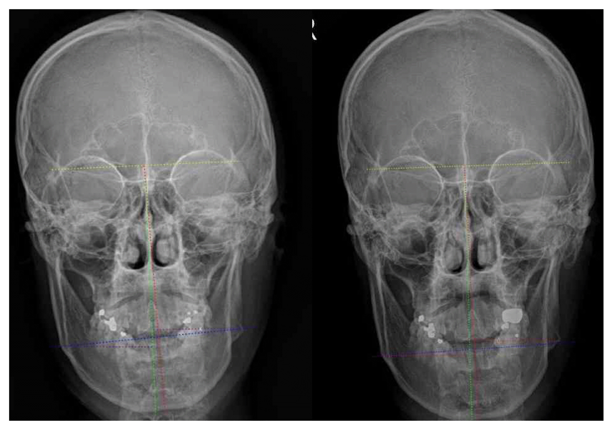
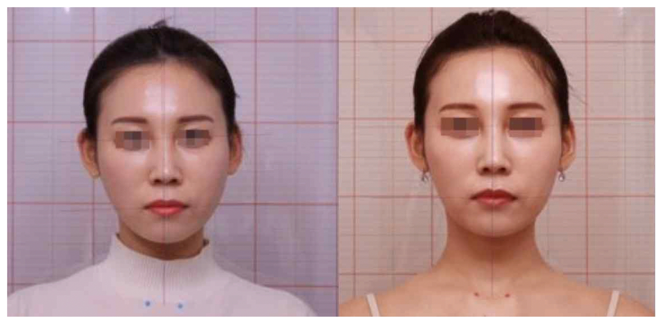
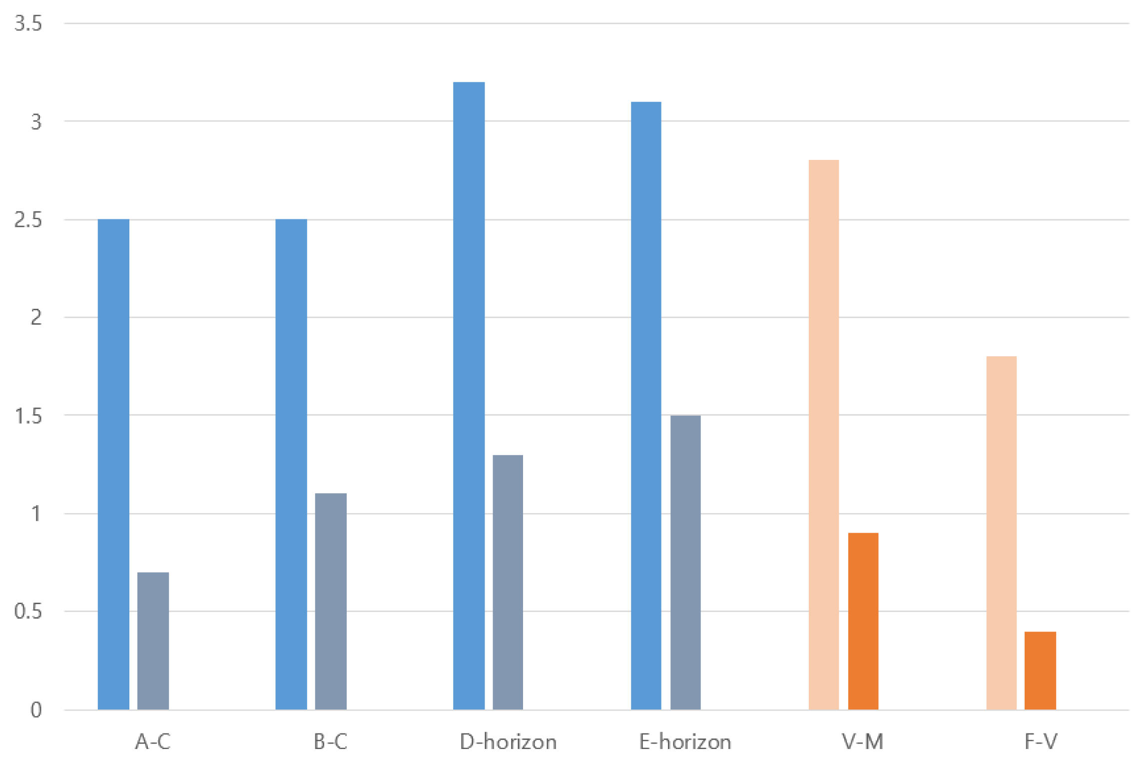
To quantify both soft and hard tissues to confirm the results of Korean medicine treatment of facial asymmetry, soft tissues quantitatively measure the displacement of the face, the slope of the left and right eyes, and the slope of the lip in order to grasp the positional displacement of the mandible. As a result, on the average, the correction effect as measured by the angle difference between A and C is 1.8±0.57, the correction effect as measured by the angle difference between B and C is 1.4±0.89, and the angle difference between D and the horizontal plane is 1.9±0.89, and the angle difference between E and the horizontal plane is 1.9±0.89. The result of reduced angle difference between A and C means that the head position shifted from the center of the body to the unilateral side was shifted to the center. The decrease in the angle difference between B and C means the restoration of the maxillary distortion relative to the mandible. In hard tissues, numerical values were measured based on the skull standard. The average distortion of the skull was 1.9±0.67, and the distortion of the lower eye was 1.4±0.41.
Conclusion
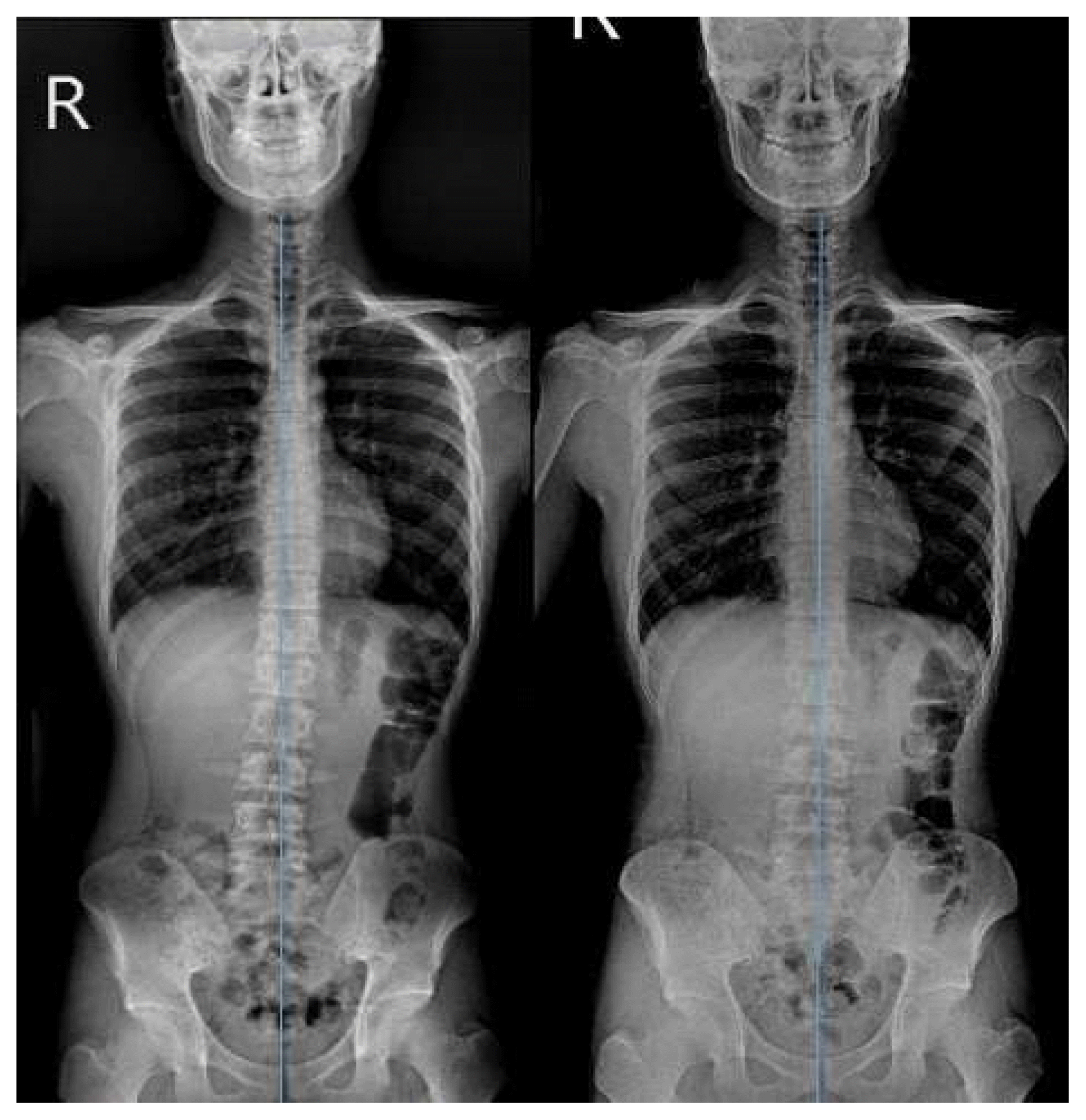
General studies on facial asymmetric treatment are limited to treatments such as surgery and orthodontics. However, this study confirmed the possibility that facial asymmetry could be corrected by Korean medical treatment consisting of reversible non-surgical treatment rather than irreversible treatment such as surgery or orthodontic treatment. In particular, Korean medicine treatment is effective for muscular asymmetry, soft asymmetry, functional asymmetry, etc. The facial asymmetric treatment of Korean medicine is not limited to the face-centered correction, but the asymmetry of the whole body may be corrected as well.
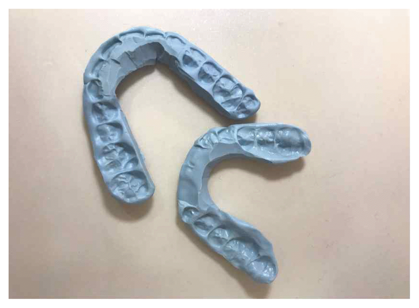
Yinyang Balance Appliance
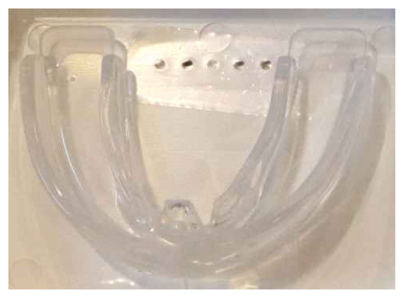
Standard Balance Appliance: IBA

Massester technique
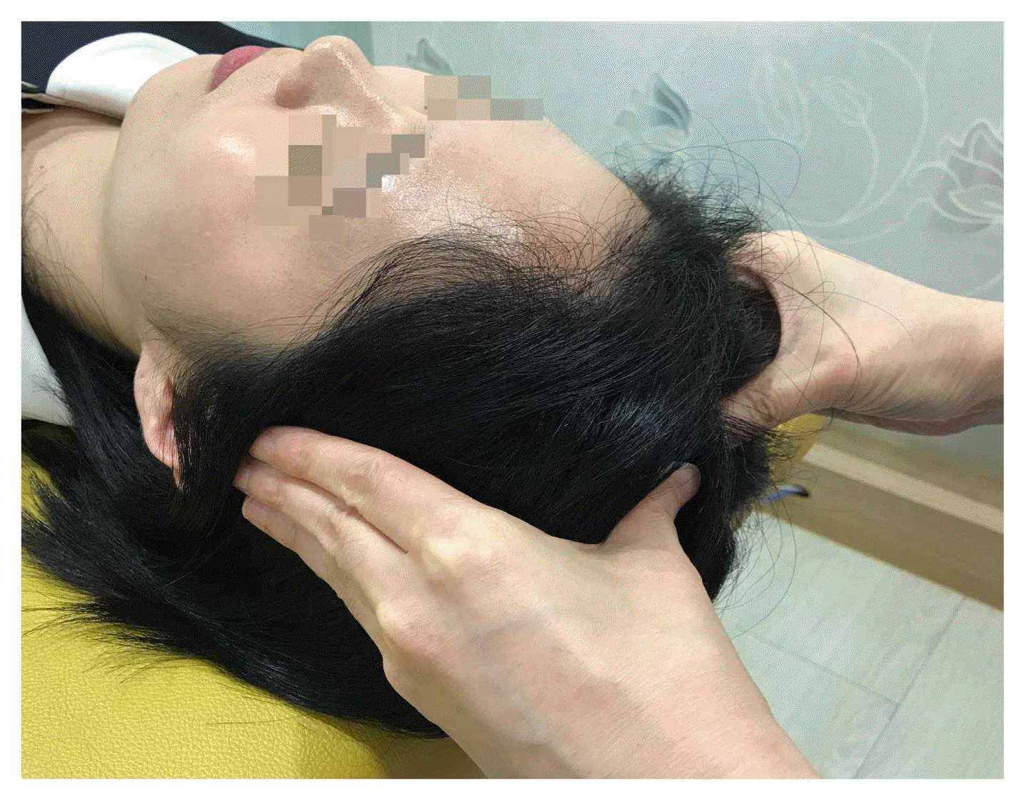
Temporalis technique

Pterygoid techniques
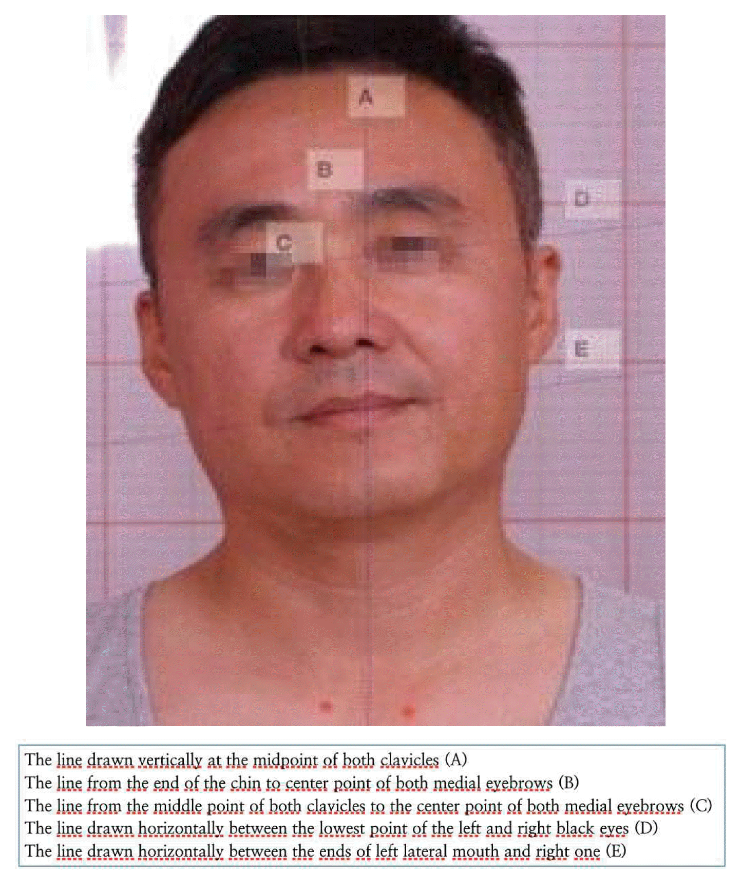
Photography analysis
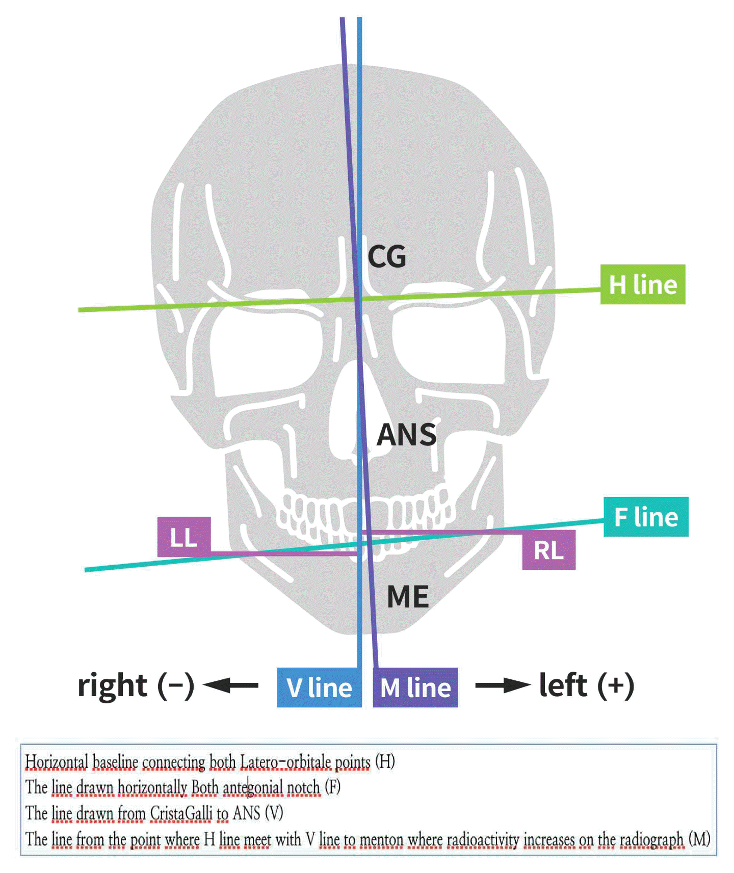
chephalography analysis

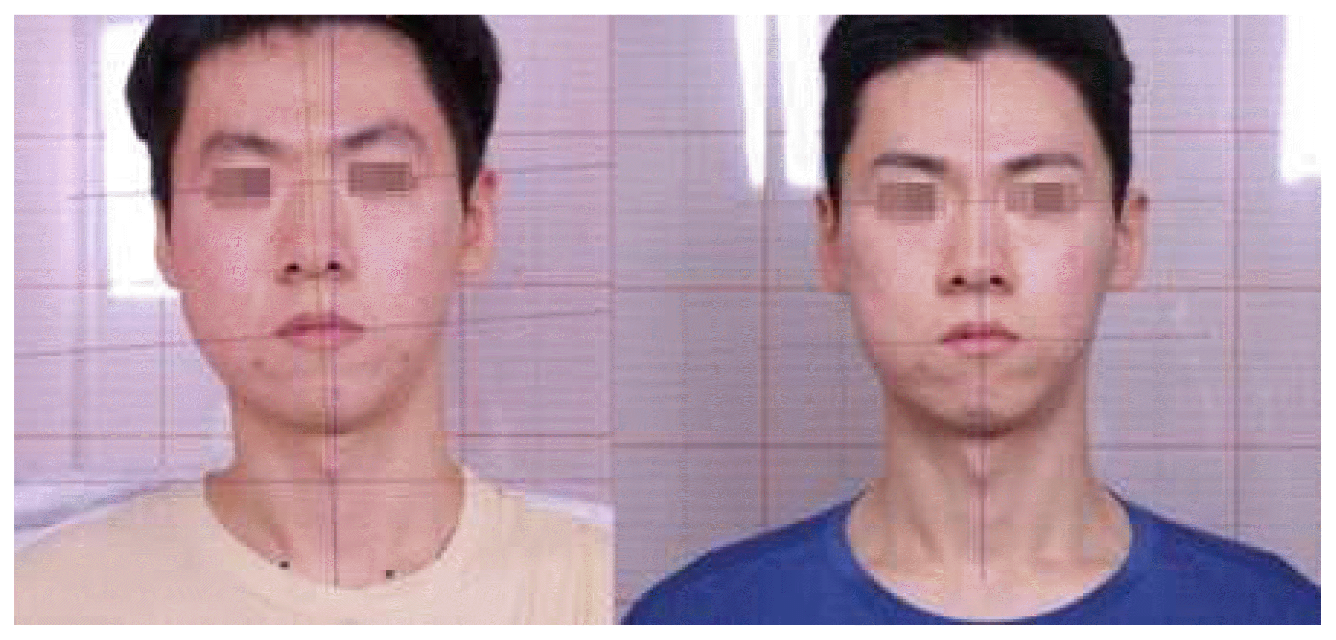
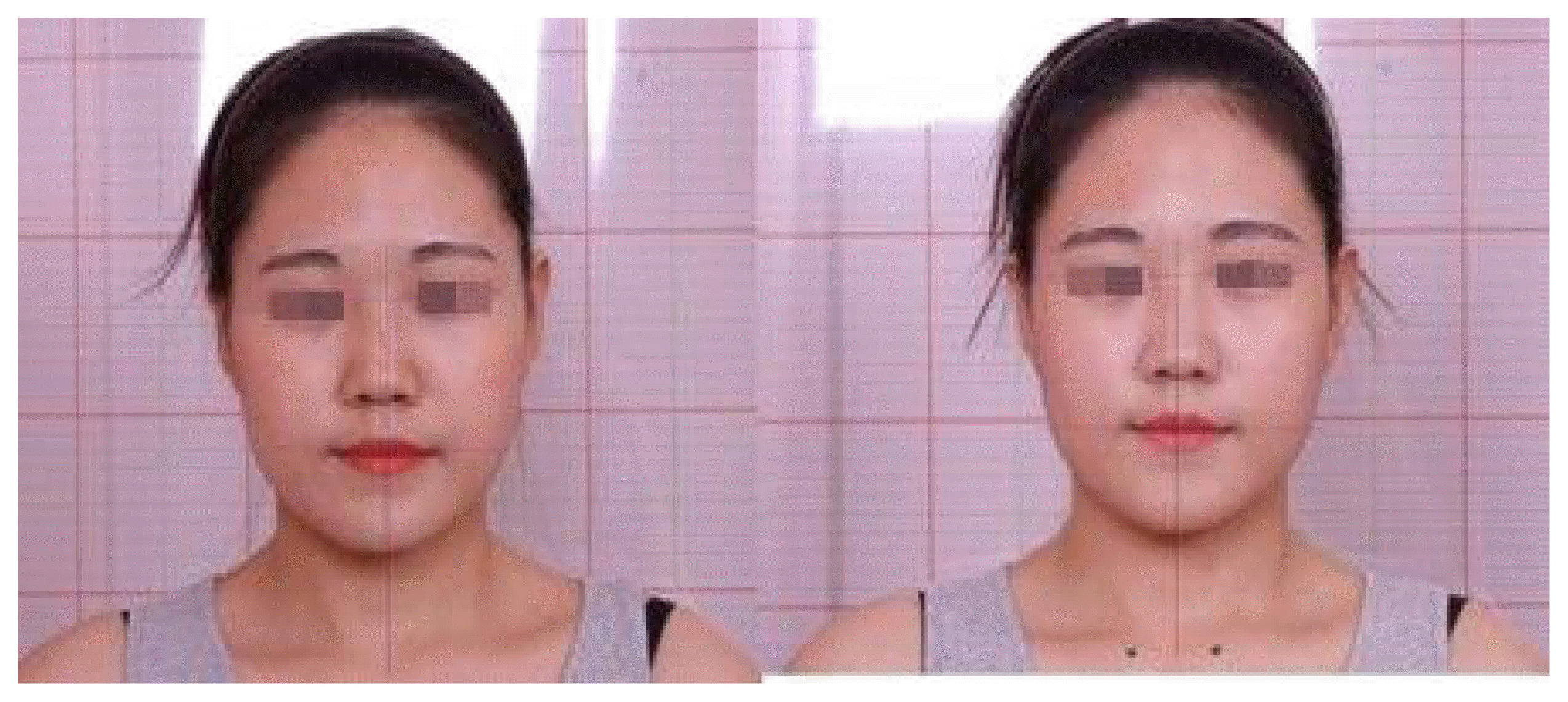
Before and after photos of Case 1
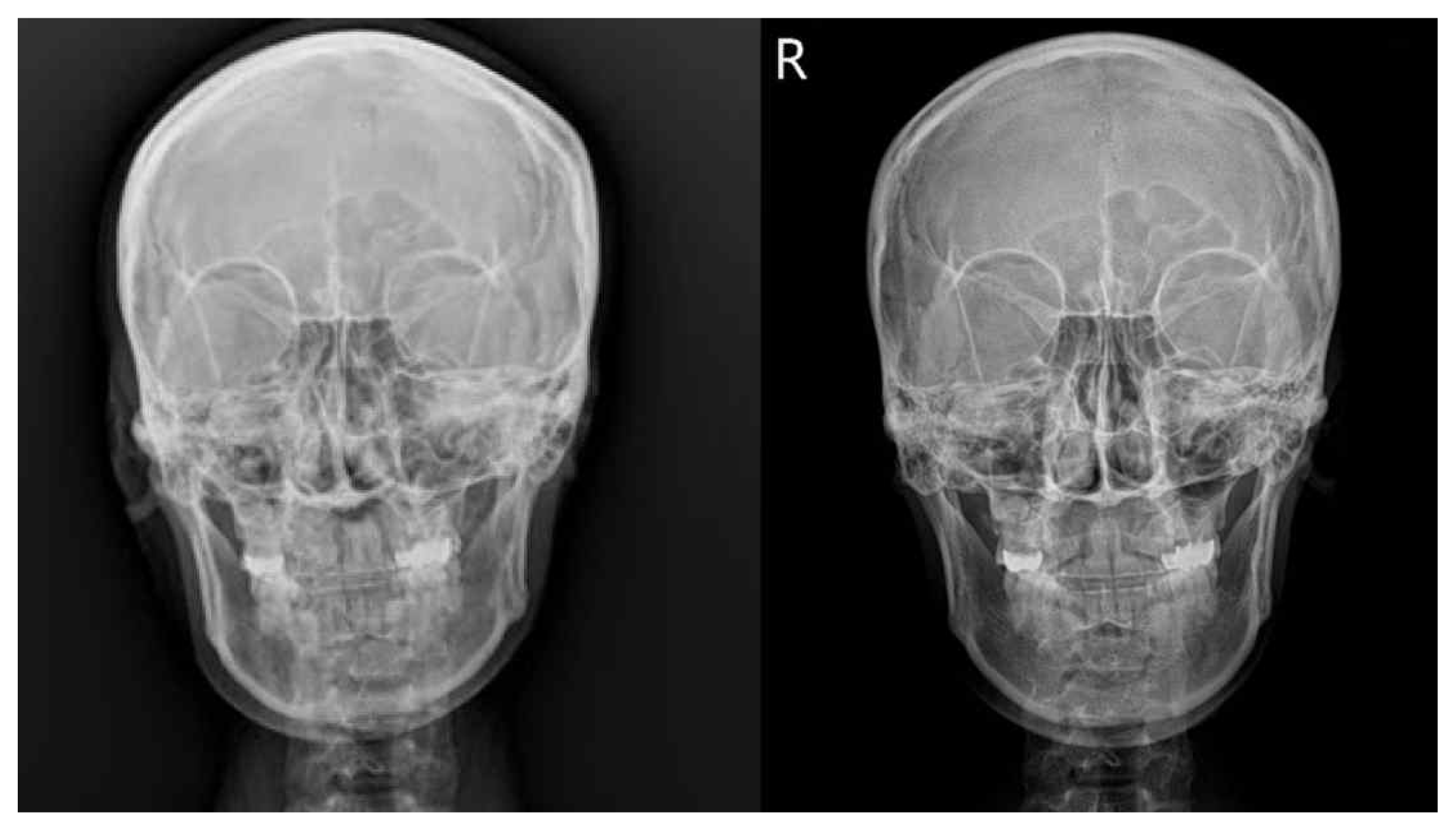
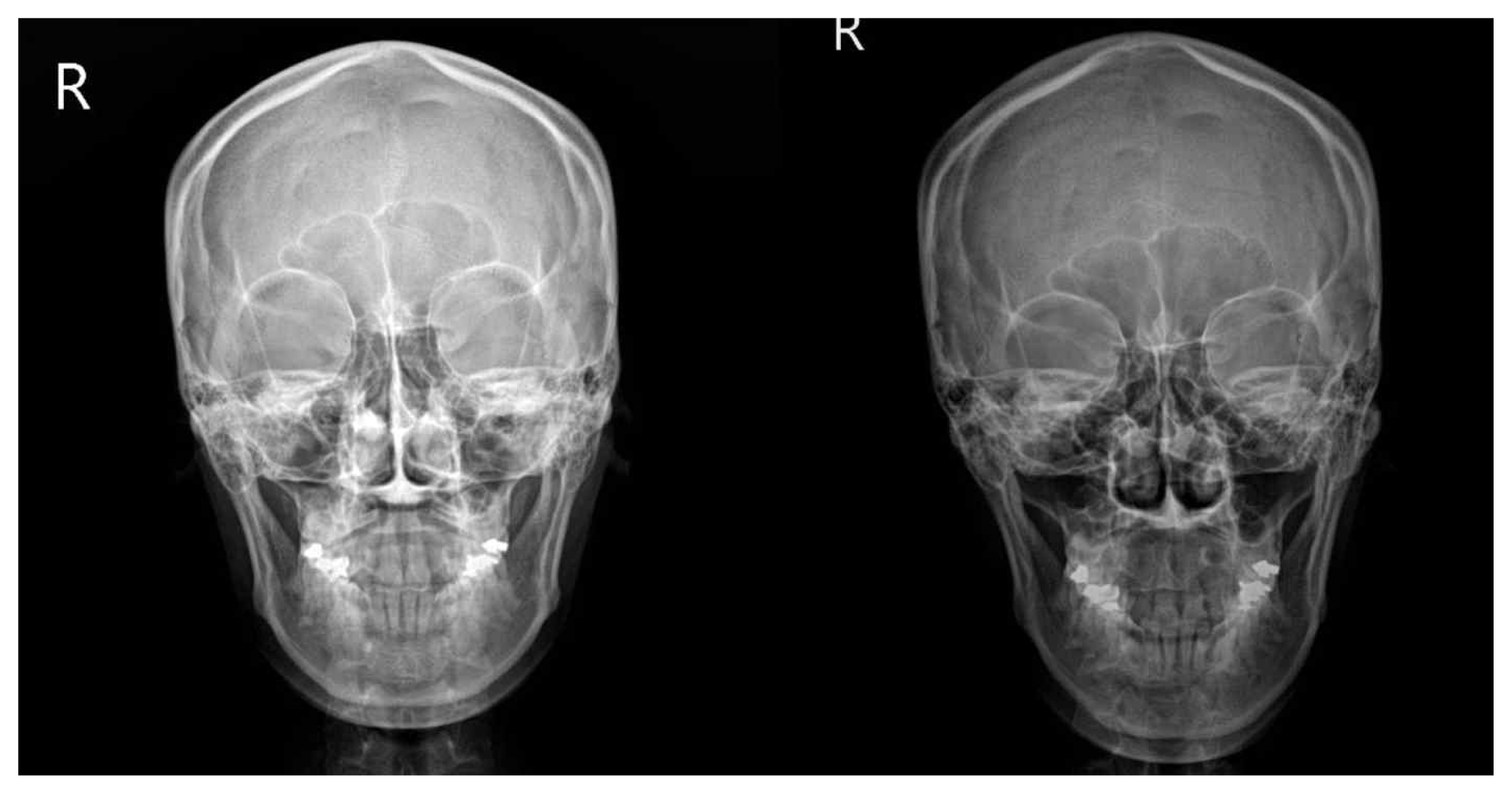
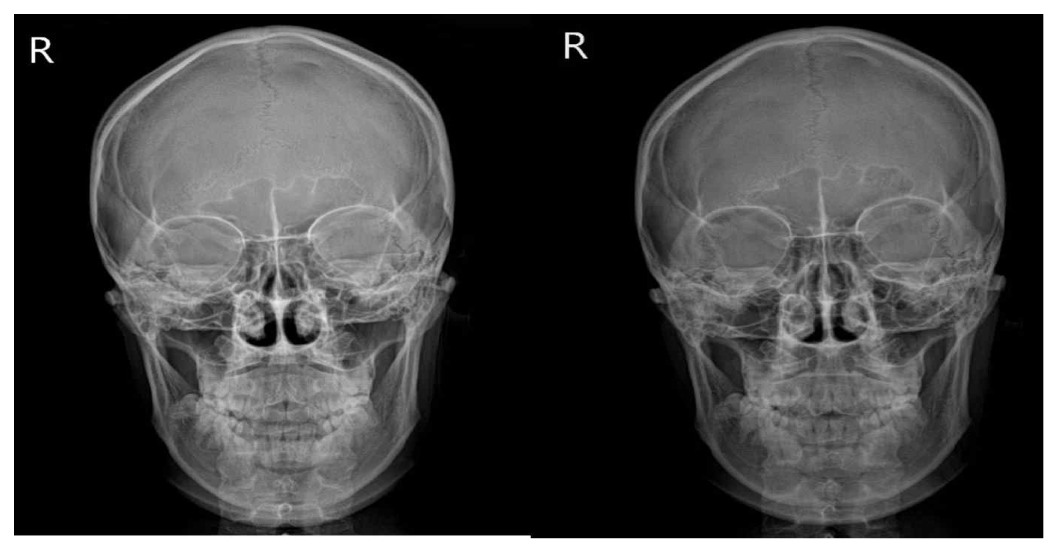
Before and after radiograms of Case 1
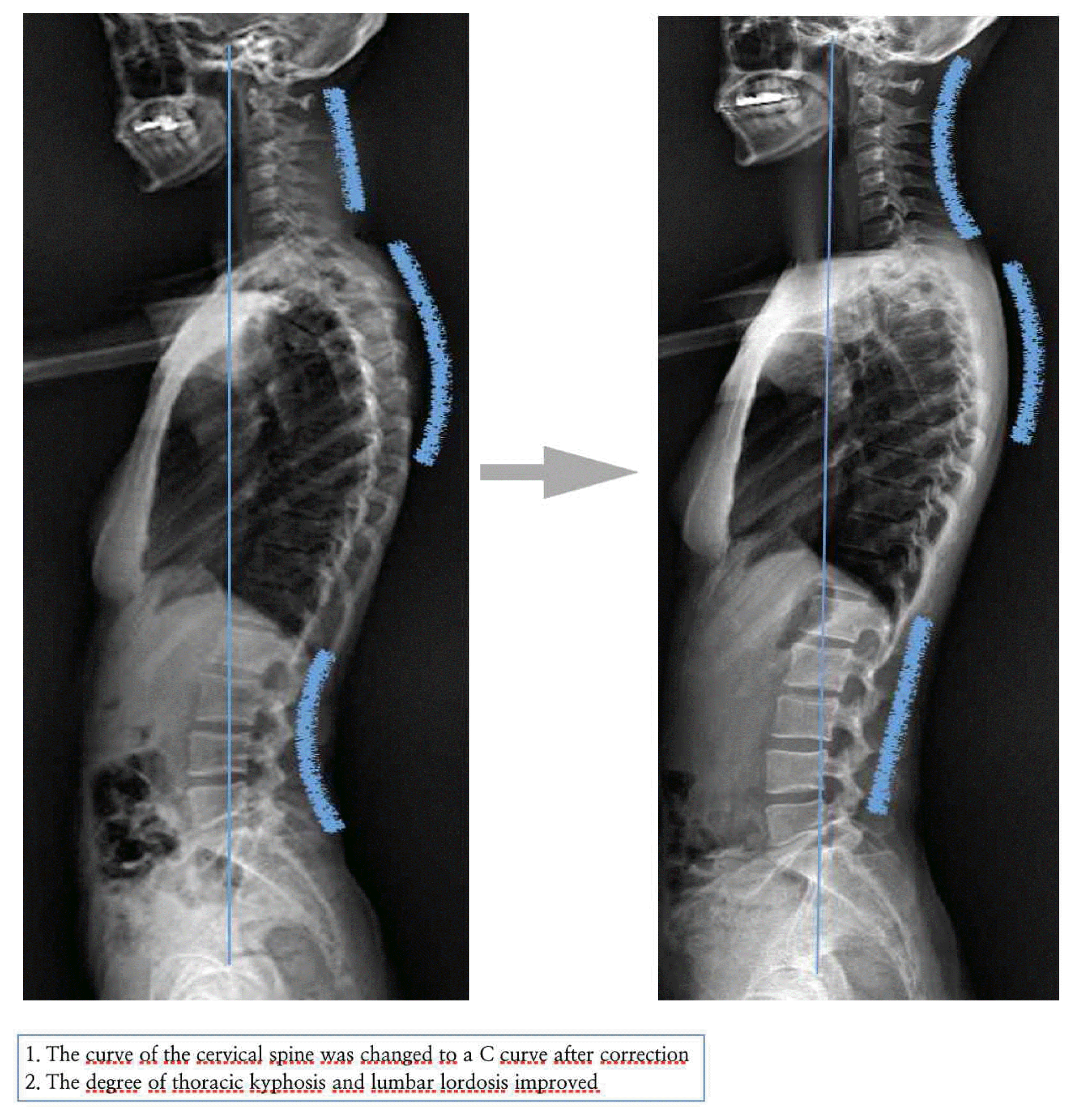
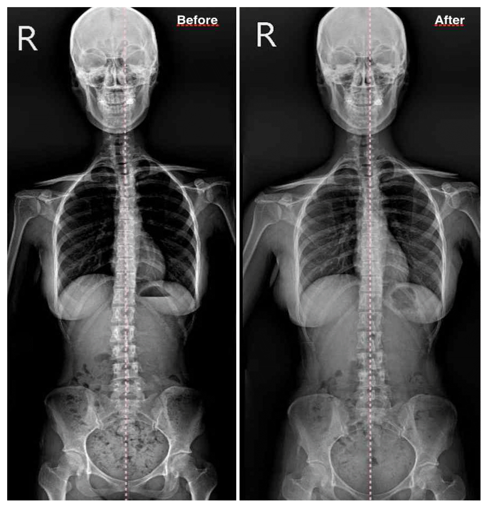
Before and after body radiograms of Case 1
Before and after photos of Case 2
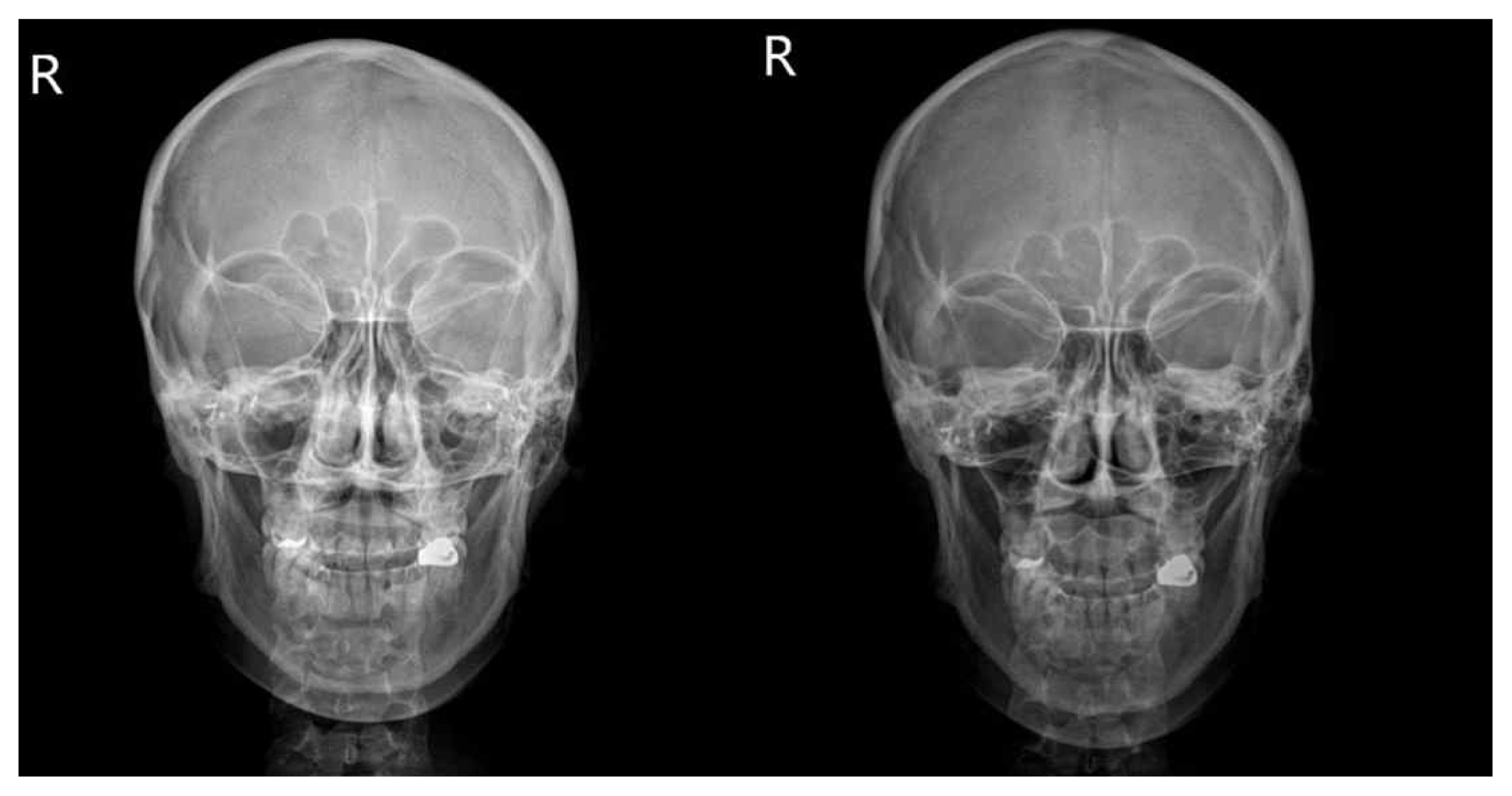
Before and after radiograms of Case 2
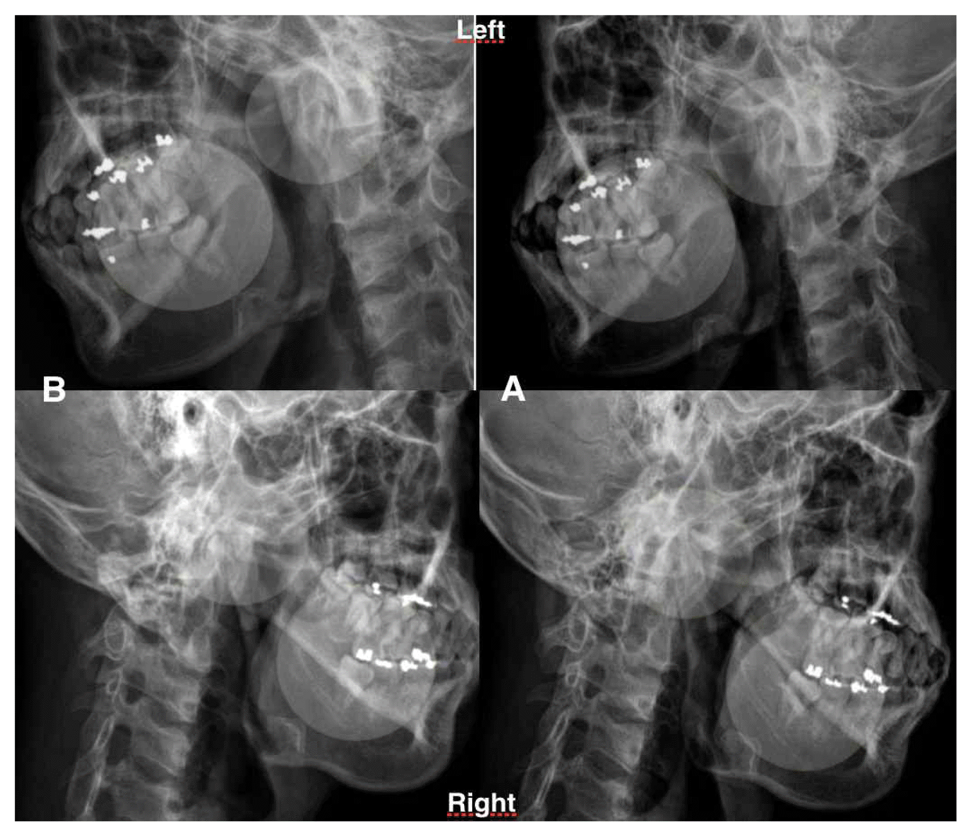
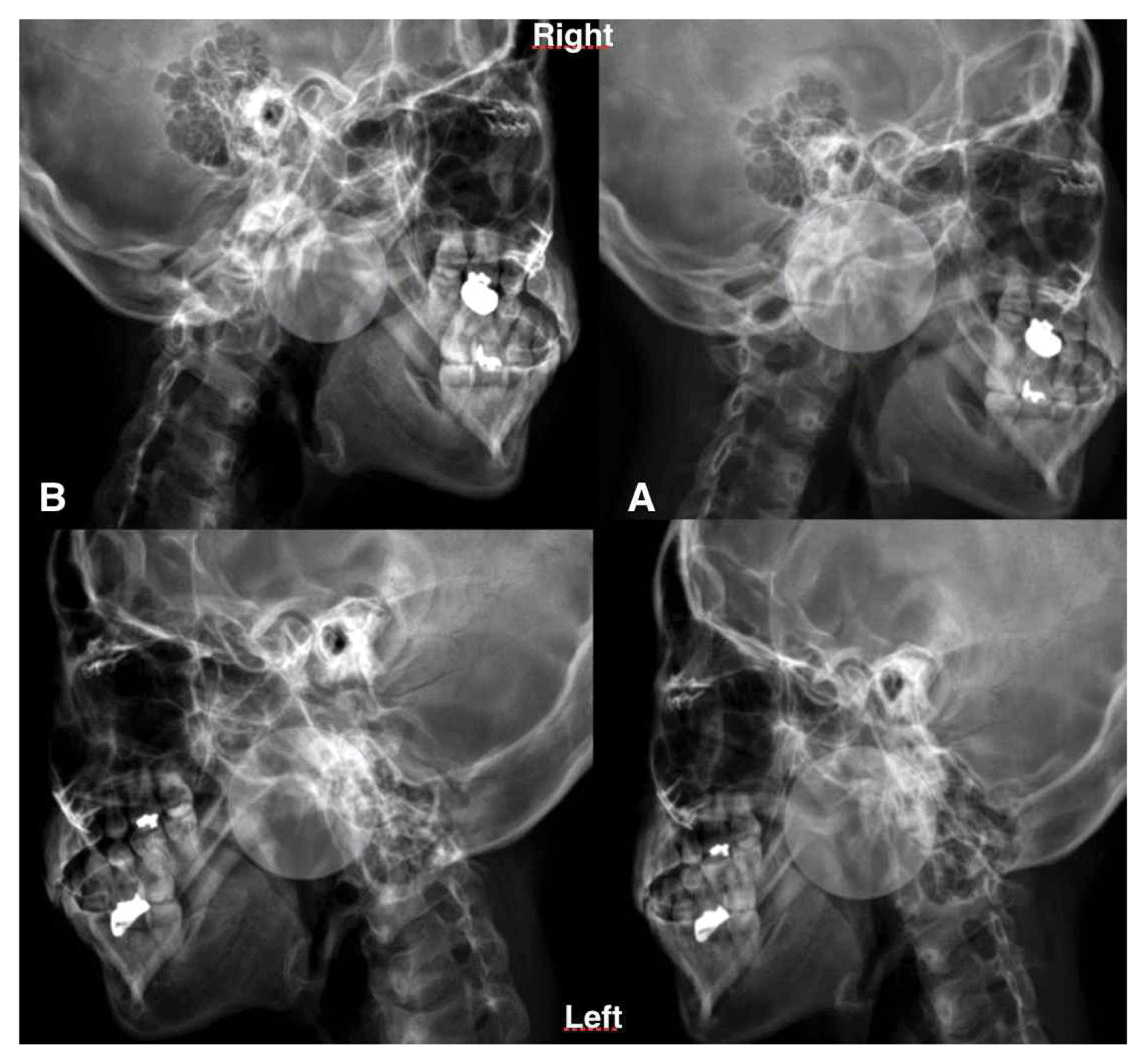
Before and after right & left lateral radiograms of Case 2
Before and after body radiograms of Case 2
Before and after photos of Case 3
Before and after radiograms of Case 3

Before and after body radiograms of Case 3

Before and after photos of Case 4
Before and after radiograms of Case 4
Before and after left lateral radiograms of Case 3
Before and after body radiograms of Case 4
Before and after photos of Case 5
Before and after radiograms of Case 5

Before and after body radiograms of Case 5
The changes of angles between lines

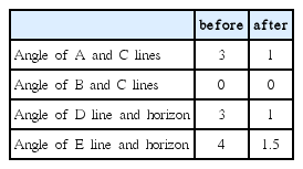
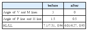
Angle before and after the procedure of Case 1
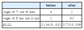
Angle before and after the procedure of Case 1
Angle before and after the procedure of Case 2
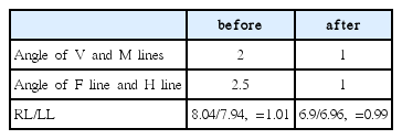
Angle before and after the procedure of Case 2
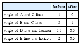
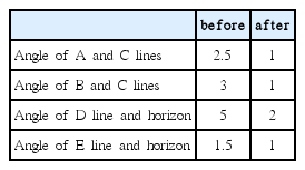
Angle before and after the procedure of Case 3
Angle before and after the procedure of Case 3

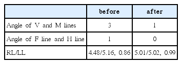
Angle before and after the procedure of Case 4
Angle before and after the procedure of Case 4
Angle before and after the procedure of Case 5
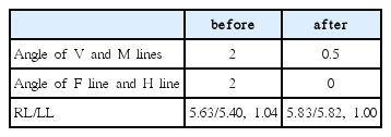
Angle before and after the procedure of Case 5
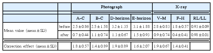
The changes of Mean value and Correction effect
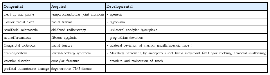
Facial Asymmetry Classification 1
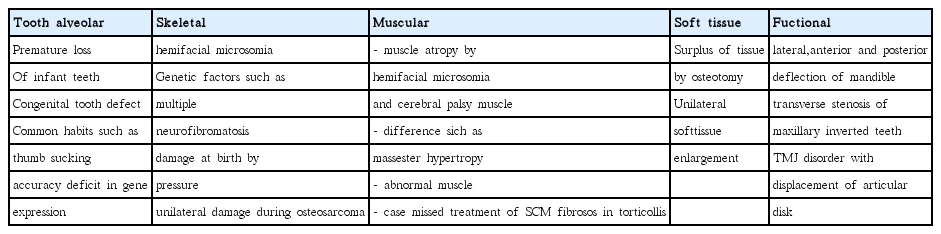
Facial Asymmetry Classification 2