Regulation of Gastric Acid Secretion of Liriope platyphylla Extract in Gastroesophageal Reflux Disease
Article information
Abstract
Objectives
The purpose of this study was to confirm the effects of Liriope platyphylla extract on relieving Gastroesophageal reflux disease (GERD) through regulation of acid secretion.
Methods
8-week-old ICR mice were divided into untreated control group (Ctrl), GERD elecitation group (GERDE), Omeprazole administrate group before GERD elicitation (OMA), and Liriope platyphylla extract administrate group before GERD elicitation (LPA). After inducing GERD, gross observation and histological examination were performed and ATP6V1B1 (ATPase H+ Transporting V1 Subunit B1), GRPR (Gastrin-releasing peptide receptor), COX-1 (Cyclooxygenase 1), 8-OHdG (8-hydroxy-2′-deoxyguanosine), Cathelicidin, p-JNK (phospho c-Jun N-terminal kinase) were observed to confirm the damage defense effect of the esophageal mucosa, acid secretion regulation, antioxidant, anti-inflammatory, mucosal protection, and apoptosis regulation
Results
OMA and LPA showed lower levels of damage compared to GERDE in gross observation and histological examination. ATP6V1B1, GRPR, and 8-OHdG showed lower positive reactions in OMA and LPA than in GERDE. COX-1 were less positive in GERDE and OMA than in Ctrl, but showed higher secretion in LPA than in Ctrl. Cathelicidin showed a decreased positive reaction in GERDE, OMA and LPA compared to Ctrl, but the decrease in positive reaction was smaller in OMA and LPA compared to GERDE. p-JNK showed increased positive reaction in GERDE, OMA and LPA than in Ctrl, but the increase in the positive reaction was smaller in the OMA and LPA compared to GERDE.
Conclusions
The effects of Liriope platyphylla extract on esophageal mucosal damage protection, acid secretion regulation, antioxidant, anti-inflammatory, mucosal protection and apoptosis regulation were confirmed.
Introduction
Gastroesophageal reflux disease (GERD) occurs when small amounts of stomach contents reflux into the esophagus, and this reflux phenomenon continues. It mainly causes symptoms such as heartburn, acid reflux, difficulty swallowing, dysphagia, nausea, and chronic cough1). This is caused by an abnormality in the sphincter that prevents the reflux of food between the stomach and the esophagus. If the damage to the esophageal mucosa persists, reflux esophagitis occurs, and when it progresses to erosive esophagitis, the mucosal damage becomes more evident. If reflux esophagitis persists for a long time, esophageal stricture may occur and as esophagitis heals, cellular dysplasia may be induced, resulting in Barrett’s esophagus. If the dysplasia of cells becomes severe, it may progress to esophageal or stomach cancer. Although the incidence of esophageal cancer isn’t relatively high, the mortality rate is very high and the prognosis is poor, so the 5-year survival rate is only less than 5%2). Therefore, if GERD is not actively treated, it may increase the potential risk factors for cancer in the long term.
Currently, the representative treatment for GERD is proton pump inhibitors (PPIs)1). PPI binds to and inactivates the proton pump of gastric parietal cells, thereby inhibiting acid secretion, which is now used as a treatment for GERD3). However, when accompanied by esophagitis and Barrett’s esophagus, PPI is used to suppress acid secretion, but this inhibition of acid secretion does not fundamentally cure or prevent esophagitis or Barrett’s esophagus2). In addition, various side effects such as increased likelihood of cardiovascular-related symptoms due to hypomagnesemia, memory impairment and neurological side effects, and the risk of toxicity due to interaction with other drugs have been reported, and the proportion of domestic refractory GERD that does not respond to drugs is approximately 26%4–6). Moreover, the use of PPIs increases the risk of osteoporotic fractures, which is pointed out as a problem in an aging society7). For this reason, research on drugs that can replace PPI by using a method different from the existing PPI mechanism is required.
Liriope platyphylla (麥門冬) is the tuber of Liriope platyphalla Wang et Tang, a perennial plant belonging to the family Liliaceae, collected in summer and dried. Liriope platyphylla is slightly cold-natured (微寒), sweet but slightly bitter flavored (甘微苦) herb. It acts on lung, stomach and heart channel (肺胃心經) and has nourishing Eum and moistening the lung (養陰潤肺), clearing away the heart-fire and relieving restlessness (淸心除煩), reinforcing the stomach and promoting production of body fluid (益胃生津) effects. It is used to treat lung dryness-cough (肺燥咳嗽), Eum deficiency-cough (陰虛咳嗽), irritability and sleeplessness (心煩失眠), hematemesis (吐血), consumptive lung disease (肺痿), lung abscess (肺癰), dysphoria with smothery sensation caused by consumptive disease (虛勞煩熱), emaciation-thirst (消渴), dry throat and mouth (咽乾口燥), constipation (便祕)8). In particular, the oriental medicinal efficacy of Liriope platyphalla, which is clearing away the heart-fire and relieving restlessness (淸心除煩), reinforcing the stomach and promoting production of body fluid (益胃生津), is thought to be applicable to the typical symptoms of GERD, such as hearburn and atypical chest pain. In addition, various studies related to Liriope platyphylla have reported anti-inflammatory and antioxidant effects9–12). Therefore, based on the anti-inflammatory and antioxidant effects reported in previous studies, Liriope platyphylla would be effective in the treatment and prevention of reflux esophagitis.
Concerning GERD and reflux esophagitis, there were studies on a variety of prescriptions and single or complex medications such as Banhasasim-tang13), Yijin-tang14,15), Jwakum-whan16), Coptis Chinensis17), Artemisia Capillaris Herba18), Evodiae Fructus19,20), Citri Pericarpium21), and Ulmus Pumila Cortex22). However, there was no study comparing the therapeutic effect of Liriope platyphylla with PPI in studies related to GERD and reflux esophagitis.
In this study, using an animal model of gastroesophageal reflux disease, the effect of Liriope platyphylla extract on the inhibition of esophageal mucosal damage was visually observed and immunohistochemical tests were performed to evaluate antioxidant, anti-inflammatory, mucosal protection, apoptosis regulation, acid secretion regulation, and visceral nervous system regulation effect of it. Through this, the inhibitory effect of Liriope platyphylla on the development of reflux esophagitis was confirmed by comparing it with Omeprazole.
Materials and Methods
1. Materials
1) Animals
8-week-old ICR male mice (Junga Bio, Korea) were acclimatized for 2 weeks in an aseptic breeding apparatus, and then mice weighing 30 ± 2 g were selected. They were divided into Untreated control group (Ctrl), Gastroesophageal reflux disease elecitation group (GERDE), Omeprazole administrate group before GERD elicitation (OMA), Liriope platyphylla extract administrate group before GERD elicitation (LPA) and 10 mice were assigned to each group.
A high-fat diet (fat 60%, carbohydrate 20%, protein 20%; DIO DIET, USA) was ad libitum to increase the frequency of reflux symptoms during the duration of the experiment23). This research process was conducted with the approval of Semyung University IACUC (IACUC number: smecae 20-09-02), and was conducted according to the NIH guidelines for the care and use of laboratory animals.
2) Experimental drug
Liriope platyphylla (100 g, Omniherb, Korea) was added to 1000 ml of distilled water, boiled for 3 hours, and then filtered. The filtrate was decompressed and concentrated to 50 ml using a rotary evaporator, and then freeze-dried to extract 24.5 g (yield: 24.5 %). After diluting the obtained Liriope platyphylla extract in physiological saline at an amount of 40 mg/kg daily from 14 days before GERD induction, 100 μl was orally administered, and the last drug administration was performed 1 hour before surgery.
Omeprazole (Shinil Pharmaceutical, Korea), a proton pump inhibitor, was used as a control drug, and 40 mg/kg was orally administered daily from 14 days before GERD induction.
2. Methods
1) Reflux esophagitis induction
Reflux esophagitis was induced using the method of Nakamura et al24). After anesthetizing the rats, which had been fasted for 24 hours, with sodium pentobarbital, the abdominal cavity was opened, and the base and cardiac region of the stomach were ligated with a micro-cable tie (Daihan Science, Korea). After 48 hours of suture, the gastroesophageal junction (GEJ) of the experimental animal was cut off and used in the experiment. In addition, to observe changes in the external shape around the gastroesophageal junction, images were taken at 4x magnification using a steroscope (Olympus, Japan).
2) Tissue section production
Cardiac perfusion was fixed with vascular rinse and 10% neutral buffered formalin (NBF). The gastroesophageal junction and lower esophagus were excised and fixed with 10% NBF at room temperature for 24 hours. After the fixed tissue was embedded in paraffin, it was prepared as a continuous section with a thickness of 5 μm.
3) Histochemical test
A phloxine-tartrazine staining method was performed to observe tissue damage to the gastric mucosa and lower esophagus at the gastroesophageal junction. After nuclear staining with Mayer’s hematoxylin for 5 minutes, it was reacted with phloxine solution for 30 minutes. After fractionation from the tartrazine solution, it was observed using an optical microscope (BX 60, Olympus, Japan).
4) Immunohistochemical test
To investigate immunohistochemical changes in the gastric mucosa of the gastroesophageal junction and the tissues of the lower esophagus, immunohistochemical staining using ATP6V1B1 (ATPase H+ Transporting V1 Subunit B1) (Abcam, USA), GRPR (Gastrin-releasing peptide receptor) (Abcam, USA), 8-OHdG (8-hydroxy-2′-deoxyguanosine) (Abcam, USA), COX-1 (Cyclooxygenase-1) (Abcam, USA), Cathelicidin (Abcam, USA) and p-JNK (phospho c-Jun N-terminal kinase) (Abcam, USA) was performed. The tissue sections were subjected to proteolysis in proteinase K (20 μg/ml) for 5 minutes, and then reacted in 10% normal goat serum, a blocking serum, for 2 hours. After reacting the primary antibody in a humidified chamber at 4°C for 72 hours, the secondary antibody, biotinylated goat anti-mouse IgG (1:100, Abcam, USA), was linked at room temperature for 24 hours and reacted with avidin biotin complex kit (Vector Lab, USA) for 1 hour at room temperature. After color development in 0.05M tris-HCl buffer (pH 7.4) containing 0.05% 3,3′-diaminobenzidine and 0.01% HCl, counterstaining was performed with hematoxylin.
5) Image analysis
Image analysis was quantified (means ± standard error) using Image Pro (Media cybernetics, USA). After magnifying the gastroesophageal connection and lower esophageal periphery by 100 times, the intensity (80–100) was measured and counted in the 20,000,000 pixel cell area.
6) Statistics
For statistics, SPSS software (SPSS 25, SPSS Inc., USA) was used, and one-way ANOVA analysis was performed. Significance was set when the P value was less than 0.05, and Tukey HSD was performed for post-test verification.
Results
1. Inhibition of gastroesophageal mucosal damage
After gastric ligation, severe hemorrhagic erosion was observed in the gastric mucosa around the gastroesophageal junction of GERDE. In contrast, a low distribution of hemorrhagic abrasions was observed in OMA and LPA. The degree of damage increased in the order of LPA, OMA, and GERDE (Fig. 1. GS-junction). As a result of histological examination through phloxine-tartrazine staining, severe mucosal damage was observed at the abrasion site. In the order of LPA, OMA, and GERDE, the injury site increased (Fig. 1. Stomach).
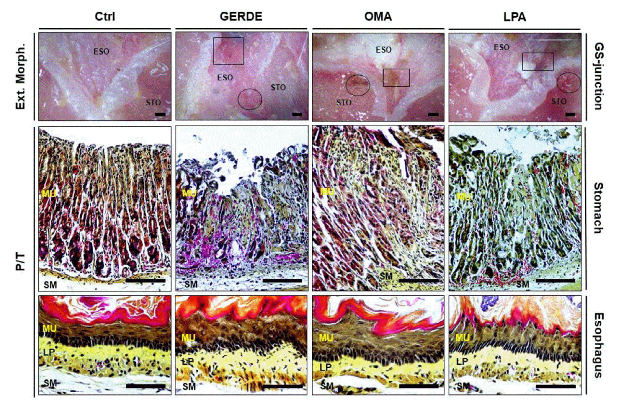
The alleviation of Gastroesophageal reflux disease (GERD) induced mucosal damages by Omeprazole and Liriope platyphylla.
Gross observation of gastroesophageal junction was performed and histological examination of stomach & esophagus abrasion site through phloxine-tartrazine staining was observed. A low distribution of hemorrhagic abrasions was observed in OMA and LPA. Histological examination through phloxine-tartrazine staining, severe mucosal damage was observed at the abrasion site. In the order of LPA, OMA, and GERDE, the injury site increased both in stomach & esophagus. GERD, Gastroesophageal reflux disease; Ctrl, no GERD elicited group; GERDE, GERD elicited group; OMA, Medicine as Omeprazole administrate group before GERD elicitation; LPA, Medicine as extraction of Liriope platyphylla administrate group before GERD elicitation; Circle, hemorrhagic erosion in stomach; Square Box, erosionin esophagus; ESO, Esophagus; STO, Stomach; MU, Mucosa; LP, Laminar propria; SM, submucosa; bar size, 50μm.
After gastric ligation, erosion was observed in the esophageal mucosa around the continuation of the gastroesophageal junction of GERDE. A low distribution of abrasion was observed in OMA and LPA. The degree of damage increased in the order of LPA, OMA, and GERDE. As a result of histological examination through phloxine-tartrazine staining, damage was observed on the apical surface of the esophagus. In the order of LPA, OMA, and GERDE, the injury site increased. (Fig. 1. Esophagus).
2. ATV6V1B1 and GRPR regulation
ATV6V1B1 positive reaction was increased by 458% in GERDE (34,911 ± 1,405 /20,000,000 pixel) compared to Ctrl (6,255 ± 411 /20,000,000 pixel). The drug treatment group showed a lower increase than GERDE. Compared to Ctrl, OMA (19,179 ± 772 /20,000,000 pixel) increased by 207% and LPA (13,700 ± 653 /20,000,000 pixel) by 119%. LPA had a 29% lower positive reaction than OMA (Fig. 2. ATV6V1B1).
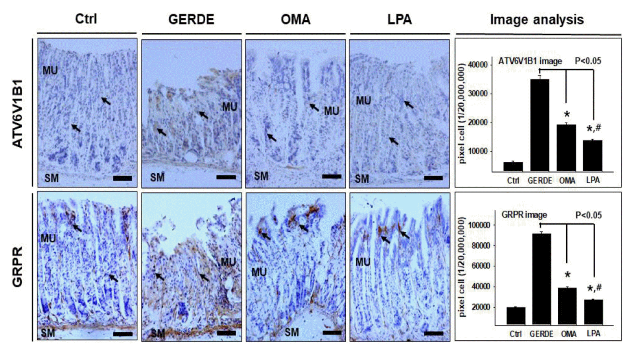
The regulation of ATPase H+ Transporting V1 Subunit B1 (ATV6V1B1) and Gastrin-releasing peptide receptor (GRPR) in stomach by Omeprazole and Liriope platyphylla.
Immunohistochemical tests of ATP6V1B1 and GRPR were performed to investigate the acid secretion regulation effect of Liriope platyphylla. Both ATP6V1B1 and GRPR showed relatively low reaction patterns in the drug-treated groups, LPA and OMA, than in the GERDE group, and the LPA group showed a lower positive reaction than the OMA group. Image analysis was quantified using Image Pro and presented as means ± standard error; *, p < 0.05 compared with GERDE; #, p < 0.05 compared with OMA; bar size, 50μm. abbrevation same as Fig. 1.
GRPR positive reaction was increased by 361% in GERDE (91,345 ± 2,187 /20,000,000 pixel) compared to Ctrl (19,792 ± 1,092 /20,000,000 pixel). The drug treatment group showed a lower increase than GERDE. Compared to Ctrl, OMA (38,869 ± 1,513 /20,000,000 pixel) increased by 96% and LPA (27,387 ± 989 /20,000,000 pixel) by 38%. LPA had a 30% lower positive reaction than OMA (Fig. 2. GRPR).
3. COX-1 inhibition
COX-1 positive reaction was reduced by 71% in GERDE (21,637 ± 1,117 /20,000,000 pixel) compared to Ctrl (73,936 ± 1,423 /20,000,000 pixel). The drug treatment group showed a lower decrease than GERDE. Compared to Ctrl, OMA (41,453 ±1,585 /20,000,000 pixel) was reduced by 44%, and LPA (52,148 ±1,463 /20,000,000 pixel) was reduced by 29%. LPA showed a 26% increase in positive reaction compared to OMA (Fig. 3.).
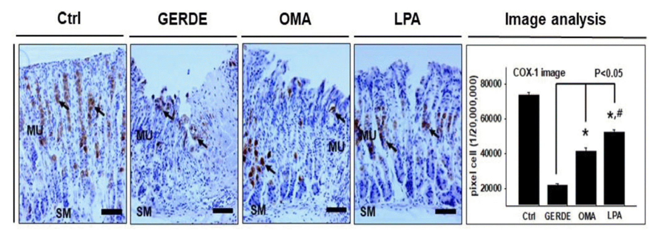
The regulation of Cyclooxygenase-1 (COX-1) in stomach by Omeprazole and Liriope platyphylla.
Immunohistochemical test of COX-1 was performed to investigate the mucosal protective effect. The COX-1 positive reaction decreased in the GERDE, OMA, and LPA compared to the Ctrl, but the decrease was smaller in the LPA than in the GERDE and OMA. Image analysis was quantified using Image Pro and presented as means ± standard error; *, p < 0.05 compared with GERDE; #, p < 0.05 compared with OMA; bar size, 50 μm. abbrevation same as Fig. 1.
4. 8-OHdG regulation
As a result of 8-OHdG immunohistochemistry to investigate damage caused by oxidative stress on the injured site, 8-OHdG positive reaction was increased by 323% in GERDE (112,410 ± 2,330 /20,000,000 pixel) compared to Ctrl (26,593 ± 1,284 /20,000,000 pixel). The drug treatment group showed a lower increase than GERDE. Compared to Ctrl, OMA (58,412 ± 1,663 /20,000,000 pixel) increased by 120% and LPA (41,790 ± 1,229 /20,000,000 pixel) by 57%. LPA showed a 28% lower increase in positive reaction than OMA (Fig. 4.).
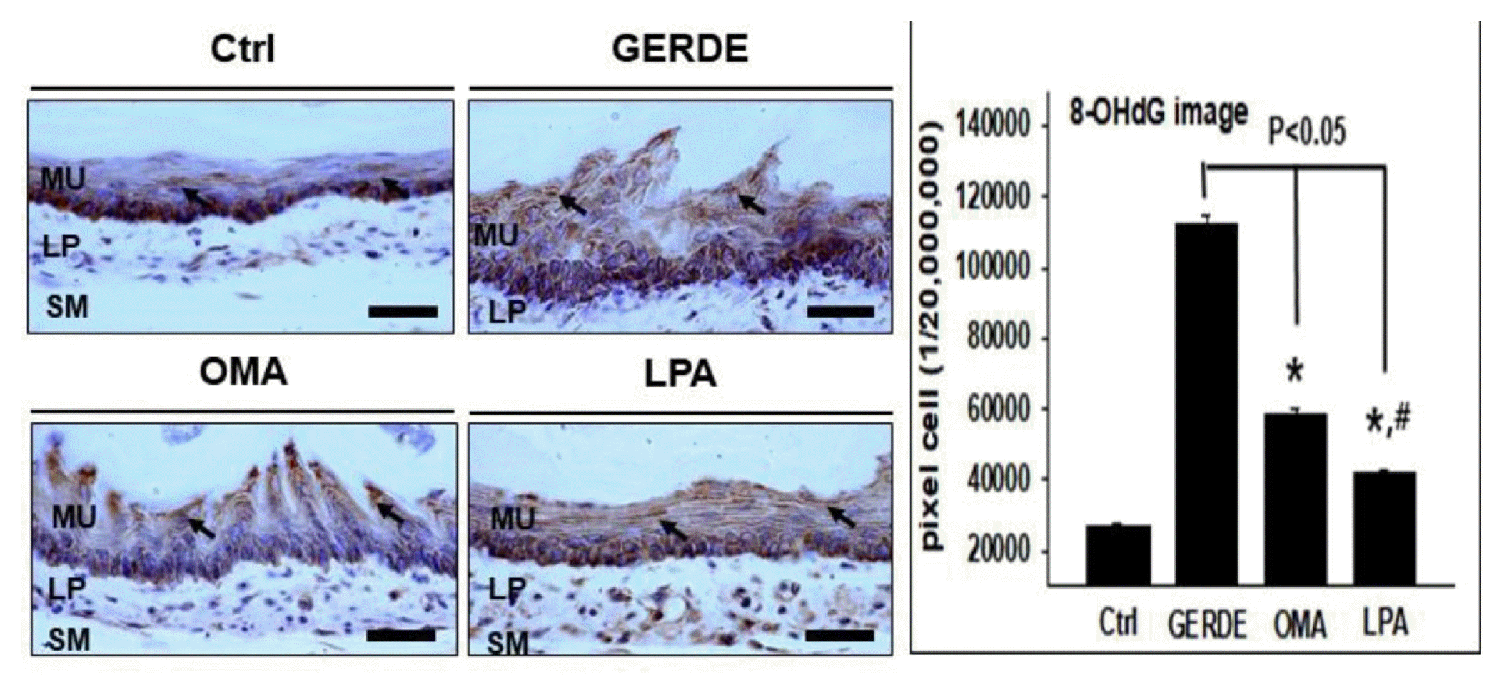
The regulation of 8-hydroxy-2′-deoxyguanosine (8-OHdG) in esophagus by Omeprazole and Liriope platyphylla.
Immunohistochemical test of 8-OHdG was performed to investigate the antioxidant effects in the gastroesophageal junction and lower esophageal region. Compared to GERDE, the drug-treated group showed a lower reaction in the order of LPA and OMA. Image analysis was quantified using Image Pro and presented as means ± standard error; *, p < 0.05 compared with GERDE; #, p < 0.05 compared with OMA; bar size, 50μm. abbrevation same as Fig. 1.
5. cathelicidin regulation
As a result of cathelicidin immunohistochemistry to investigate the formation of a protective barrier against the outside, cathelicidin positive reaction was reduced by 87% in GERDE (4,815 ± 281 /20,000,000 pixel) compared to Ctrl (36,755 ± 716 /20,000,000 pixel). The drug treatment group showed a lower decrease than GERDE. Compared to Ctrl, OMA (11,364 ± 519 /20,000,000 pixel) was reduced by 69% and LPA (23,443 ± 1,030 /20,000,000 pixel) by 36%. LPA showed a 106% higher increase in positive reaction than OMA (Fig. 5.).
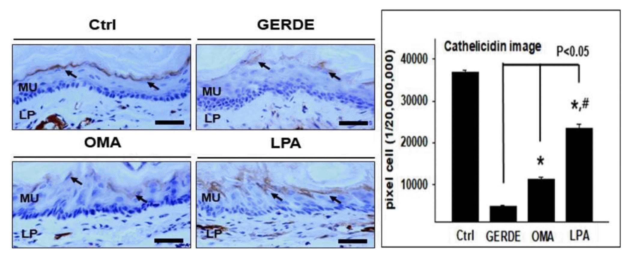
The regulation of Cathelicidin in esophagus by Omeprazole and Liriope platyphylla.
Immunohistochemical test of Cathelicidin was performed to investigate the mucosal protective effect in the lower esophageal region. The positive reaction in GERDE was decreased compared to the Ctrl, and the LPA and OMA were less decreased compared to GERDE. In addition, the reduction of the positive reaction in LPA was smaller than that in OMA. Image analysis was quantified using Image Pro and presented as means ± standard error; *, p < 0.05 compared with GERDE; #, p < 0.05 compared with OMA; bar size, 50μm. abbrevation same as Fig. 1.
6. p-JNK regulation
As a result of p-JNK immunohistochemistry to investigate the apoptosis modulating effect, the positive p-JNK reaction was increased by 695% in GERDE (44,709 ± 1,005 /20,000,000 pixel) compared to Ctrl (5,627 ± 317 /20,000,000 pixel). The drug treatment group showed a lower increase than GERDE. Compared to Ctrl, OMA (33,322 ± 906 /20,000,000 pixel) increased by 492% and LPA (18,948 ± 504 /20,000,000 pixel) by 237%. LPA showed a 43% lower increase in positive reaction than OMA (Fig. 6.).
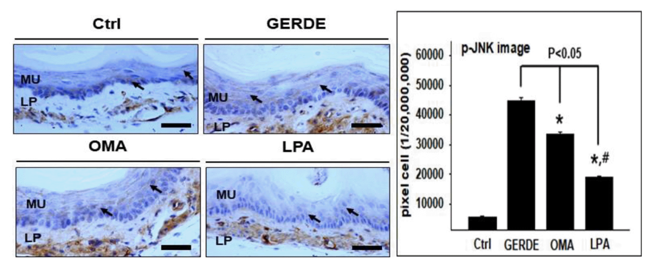
The regulation of phospho c-Jun N-terminal kinase (p-JNK) in esophagus by Omeprazole and Liriope platyphylla.
Immunohistochemical test of p-JNK was performed to investigate the effect of modulating apoptosis in the gastroesophageal junction and lower esophageal region. The positive reaction of p-JNK was increased in the GERDE compared to Ctrl, and LPA and OMA increased less than GERDE. Image analysis was quantified using Image Pro and presented as means ± standard error; *, p < 0.05 compared with GERDE; #, p < 0.05 compared with OMA; bar size, 50μm. abbrevation same as Fig. 1.
Discussion
This study investigated the inhibitory effect of Liriope platyphylla on the onset of reflux esophagitis in the gastroesophageal junction and lower esophageal region using an animal model of reflux esophagitis. The effect of inhibiting damage to the esophageal mucosa was visually observed, and antioxidant, anti-inflammatory, gastric acid secretion control, mucosal protection, and apoptosis control were observed and compared with Omeprazole. Through this, it was confirmed whether Liriope platyphylla had a superior effect compared to the conventional treatment for GERD and reflux esophagitis.
First, through gross observation at the gastroesophageal junction and lower esophageal region and histological examination through phloxine-tartrazine staining, the GERDE-induced group showed severe hemorrhagic erosion, whereas the drug treatment group showed a low degree of damage in the order of LPA and OMA.
In this study, immunohistochemical tests of ATP6V1B1 and GRPR were performed to investigate the acid secretion regulation effect of Liriope platyphylla. ATP6V1B1 is a gene that transcribes the B1 subunit of the proton pump, and is an important factor involved in maintaining acid secretion homeostasis in the body25,26). GRPR is a receptor for gastrin-releasing peptide (GRP), and when GRP is released from the enteric nervous system, the local concentration of gastrin increases to promote gastric acid secretion, and is closely related to inflammatory conditions27,28). Both ATP6V1B1 and GRPR showed relatively low reaction patterns in the drug-treated groups, LPA and OMA, than in the GERDE group, and the LPA group showed a lower positive reaction than the OMA group. These results indicate that Liriope platyphylla can inhibit acid secretion by regulating the proton pump function and gastrin secretion function, and furthermore, it shows the possibility that Liriope platyphylla can be used for various diseases caused by excessive gastric acid secretion as well as GERD.
The mucosal protective effect at the gastroesophageal junction was observed through COX-1 immunohistochemistry. COX-1 is an important enzyme in the biosynthesis process from arachidonic acid to prostaglandin, and promotes the formation of the lining of the gastric mucosa to protect the stomach and inhibits the secretion of gastric acid and pepsin29). Therefore, COX-1 can be said to be a representative gastric mucosal protective factor, and when COX-1 is deficient, gastric mucosal damage occurs primarily30). The COX-1 positive reaction decreased in the GERDE, OMA, and LPA compared to the Ctrl, but the decrease was smaller in the LPA than in the GERDE and OMA. The smaller decrease in positive reaction in the LPA indicates the possibility that Liriope platyphylla may have a direct effect on gastric mucosal protection and damage prevention.
Antioxidant effects in the gastroesophageal junction and lower esophageal region were observed by 8-OHdG immunohistochemistry. 8-OHdG is formed when reactive oxygen species (NOS) attack guanine at the dinucleotide site of somatic cells, and is an important biomarker indicating the degree of oxidative stress damage31). In general, an increase in 8-OHdG not only means that the degree of oxidative damage has increased, but it is also widely used to measure risk factors for various diseases, including cancer32). As a result of the test, compared to GERDE, the drug-treated group showed a lower reaction in the order of LPA and OMA. This means that Liriope platyphylla has an antioxidant effect by inhibiting damage caused by free radicals, and that its effect may be superior to that of Omeprazole.
The mucosal protective effect in the lower esophageal region was observed by cathelicidin immunohistochemistry. Cathelicidin is an antibacterial peptide and plays an important role in human immunity by forming a protective barrier against external stimuli on the mucosal surface of the gastrointestinal tract33,34). As a result of the study, the positive reaction in the GERDE group was decreased compared to the Ctrl group, and the LPA group and OMA group were less decreased compared to the GERDE group. In addition, the reduction of the positive reaction in the LPA group was smaller than that in the OMA group, demonstrating that Liriope platyphylla had a mucosal protective effect by promoting the formation of the gastric mucosal lining, enhancing the gastric mucosal protective effect, and improving the immune capacity of the mucosal surface.
The effect of modulating apoptosis in the gastroesophageal junction and lower esophageal region was observed by p-JNK immunohistochemistry. P-JNK is a phosphorylated JNK, associated with an increase in apoptosis, is involved in cell apoptosis and inflammatory reactions in reaction to stress stimuli35–37). In this study, the positive reaction of p-JNK was increased in the GERDE group compared to the Ctrl group, and the LPA group and OMA group increased less than the GERDE group. Therefore, it is shown that Liriope platyphylla is superior to Omeprazole in the degree of apoptosis control effect and it is thought that Liriope platyphylla was involved in signaling and mediating processes for apoptosis, thus inhibiting apoptosis.
As such, at the gastroesophageal junction and lower esophageal region, Liriope platyphylla showed more significant results than Omeprazole in inhibiting damage to the esophageal mucosa, controlling antioxidant, acid secretion, anti-inflammatory, mucosal protection, and controlling apoptosis. Based on these research results, it is expected that Liriope platyphylla will have a sufficiently high potential for use in promoting the formation of the gastric mucosa lining, inhibiting gastric mucosal damage caused by gastric acid, and inhibiting inflammation and apoptosis. Research is needed to explain the mechanisms for GERD treatment and prevention of Liriope platyphylla extracts in the future, and further research on safety and validation of Liriope platyphylla is thought to be necessary.
Conclusion
In this study, the effects of Liriope platyphylla on esophageal mucosa damage defense, acid secretion regulation, antioxidant, anti-inflammatory, mucosal protection, and apoptosis regulation were confirmed through reflux esophagitis animal model experiments.
Gross observation and histological examination confirmed the damage defense effect of the esophageal mucosa of Liriope platyphyla.
ATP6V1B1 and GRPR immunohistochemical tests confirmed the acid secretion regulation effect of Liriope platyphyla.
The protective effect of Liriope platyphyla on gastric mucosa was confirmed through COX-1 immunohistochemistry.
The antioxidant effect of Liriope platyphyla was confirmed through 8-OHdG immunohistochemistry.
The protective effect of Liriope platyphyla on the lower esophageal mucosa was confirmed by cathelicidin immunohistochemistry.
The apoptosis-regulating effect of Liriope platyphyla was confirmed through p-JNK immunohistochemistry.
Acknowledgement
This study was supported by the clinical research fund of Pusan National University Hospital in 2021.
