A Case Report of Ultrasound-guided Bee Venom Pharmacopuncture on Plantar Fasciitis

Article information

J Korean Med. 2023;44(1):108-116
Publication date (electronic) : 2023 March 1
doi : https://doi.org/10.13048/jkm.23010
1Dept. of Sasang Constitutional Medicine, Woosuk University Korean Medicine Hospital
Correspondence to: Soo-Jung Park, Dept. of Sasang Constitutional Medicine, Woosuk University Korean Medicine Hospital, 46 Eoeun-ro, Wansan-gu, Jeonju-si, Jeollabuk-do, Republic of Korea 54987, Tel: +82-63-220-8676, Fax: +82-63-291-9312, E-mail: taorgi@hanmail.net
Received 2023 January 16; Revised 2023 February 8; Accepted 2023 February 14.

Abstract

Objectives

This study is to report the usefulness of ultrasound in diagnosing plantar fasciitis and the effectiveness of ultrasound-guided bee venom pharmacopuncture as a treatment of it.

Methods

A 61-year-old woman suffered from plantar fasciitis on her right foot for 1 month. The sonographic findings were fascial thickening, blurring of perifascial border and perifascial effusion. The ultrasound-guided bee venom pharmacopuncture for 6 times and conventional Korean medicine therapies like acupuncture and moxibustion for 12 times in 5 weeks. Numeric rating scale (NRS) and foot function index(FFI) was used to evaluate the pain and its progress. and thickness of plantar fascia was measured by sonography every week.

Results

After treatments, NRS and FFI were reduced from 10 to 1 and from 190 to 72. The thickness of fascia was reduced from 0.43cm to 0.40. Blurring and effusion of perifascial border were also improved.

Conclusion

This report suggests that the Ultrasound-guided Bee Venom Pharmacopuncture is effective for Plantar Fasciitis. Ultrasonography could be one of the most valuable items in the clinical practice of Korean medicine doctors who seek minimally invasive treatment.

Fig. 1

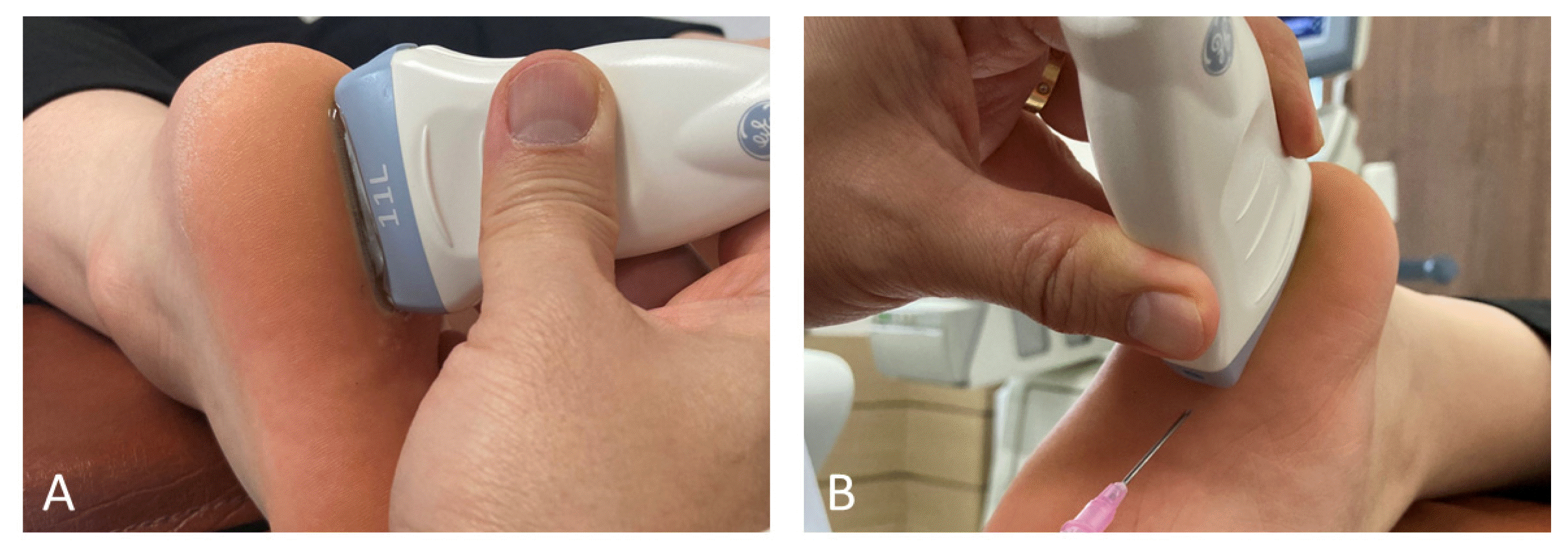
(A) Ultrasound scanning posture and transducer position of the plantar fascia. (B) Ultrasound-guided pharmacopuncture. The direction of needle insertion(Example).

Fig. 1

A 61-year-old woman with plantar fasciitis on her right foot.

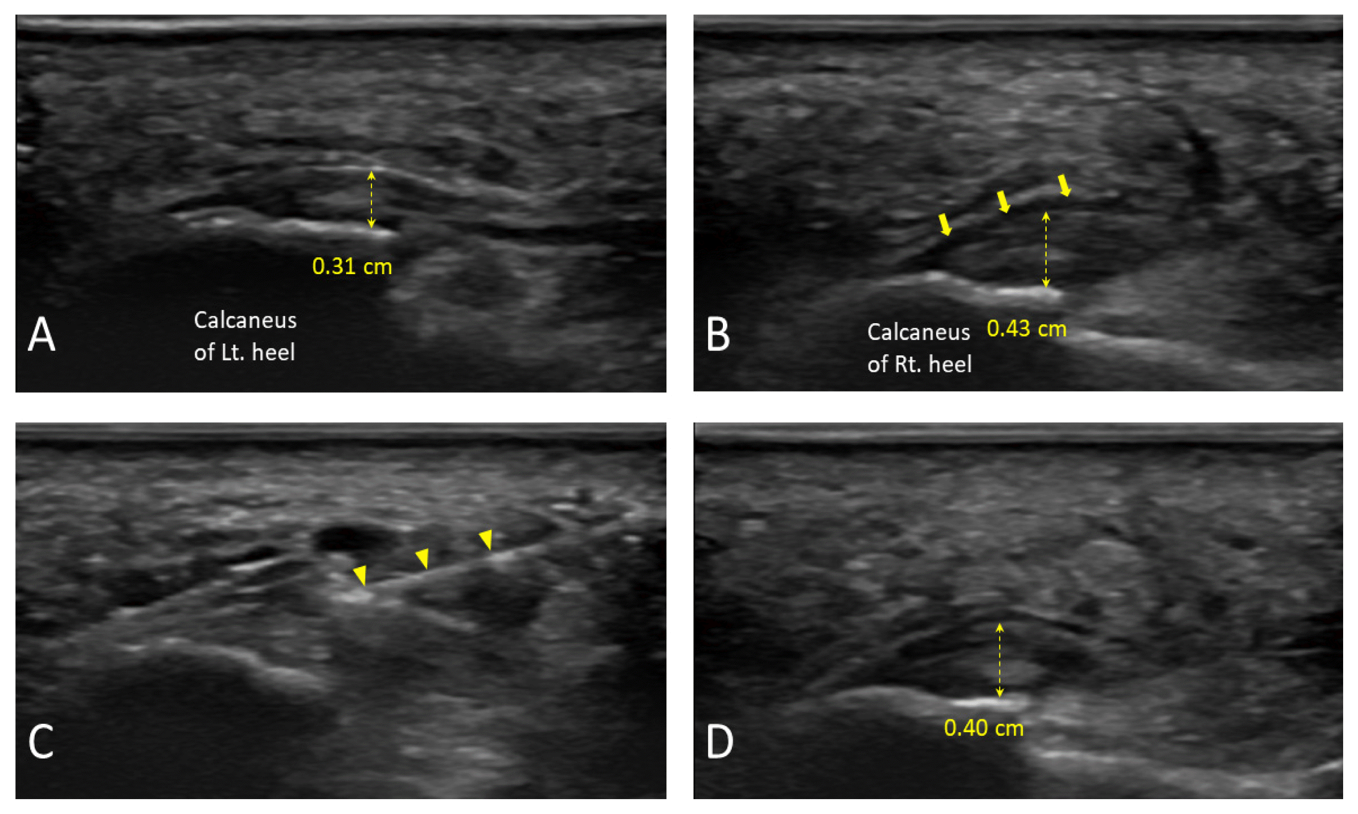
(A) Long-axis ultrasound image of the plantar fascia on the normal left calcaneus. Its thickness was 0.31cm. (B) Sonographic findings of plantar fasciitis on right calcaneus before treatment. It shows fascial thickening of 0.43cm, hypoechogenicity, blurring of perifascial border, and perifascial effusion(arrows). (C) Ultrasound-guided pharmacopuncturetherapy. The needle(arrowheads) was inserted obliquely from anterior to posterior to reach the outer border of the fascia and sweet bee venom was injected. (D) After treatments of about 1 month, fascial thickening was reduced to 0.40cm and perifascial effusions were improved.

Timeline of Ultrasound-guided Bee Venom Pharmacopuncture and Korean medicine treatment with Changings in Pain and Symptom Evaluation Indicators of Plantar Fasciitis

References

1. Buchbinder R. 2004;Plantar fasciitis. N Engl J Med 350(21):2159–2166. https://doi.org/10.1056/NEJMcp032745 .
2. Crawford F, Thomson C. 2003;Interventions for treating plantar heel pain. Cochrane Database Syst Rev 3:CD000416. https://doi.org/10.1002/14651858.CD000416 .
3. Koh NY, Kim CG, Ko YS, Lee JH. 2015;Acupuncture Treatment of Plantar Fasciitis: A Literature Review. J Korean Med Rehabil 25(2):97–110.
4. Ha WB. 2022;A Case Report of Talipes Cavus-Type Plantar Fasciitis Treated with Acupotomy and Fascia Chuna Therapy. The Journal of Korea Chuna Manual Medicine for Spine & Nerves 17(1):47–53. https://doi.org/10.30581/jcmm.2022.17.1.47 .
5. Hwang HJ, Lee KJ, Park YH, Keum DH. 2008;Two Clinical Cases on Plantar Fasciitis Using Myofascial Releasing Therapy and Acupuncture Therapy. J Korean Med Rehabil 18(2):111–118.
6. Kim HJ, Ji YS, Lee SM, Jeon JH, Kim YI. 2013;A Systematic Review of Clinical Study of Bee Venom Acupuncture. J Acupunct Res 30(4):151–159. https://doi.org/10.13045/acupunct.2013031 .
7. Ahn KH, Kim KH, Hwang HS, Song HS, Kwon SJ, Lee SN, et al. 2002;The Effect of Bee-venom Acupuncture on Heel Pain. The Journal of Korean Acupuncture & Moxibustion Society 19(5):149–160.
8. Lee SJ, Ki SH, Koh DK, Lee SH, Lim HH, Song YK. 2022;A Study on the Exploration of Treatment Area of Visceral Chuna Manual Therapy Using Ultrasound Image Data. J Korean Med Rehabil 32(2):139–154. https://doi.org/10.18325/jkmr.2022.32.2.139 .
9. Ahn TS, Moon JH, Park CY, Oh MJ, Choi YM. 2019;The Effectiveness of Ultrasound-Guided Essential Bee Venom Pharmacopuncture Combined with Integrative Korean Medical Treatment for Rib Fracture: A Case Study. J Korean Med Rehabil 29(3):157–163. https://doi.org/10.18325/jkmr.2019.29.3.157 .
10. Kim YH, Oh TY, Lee EJ, Oh MS. 2019;A Comparative Study on the Pain and Treatment Satisfaction between Korean Medical Treatment Combined with Ultrasound Guided Soyeom Pharmacopuncture Therapy in Thoracic Paravertebral Space and Non-Guided Soyeom Pharmacopuncture Therapy on Patients with Ribs Fracture: A Retrospective Study. J Korean Med Rehabil 29(3):103–112. https://doi.org/10.18325/jkmr.2019.29.3.103 .
11. Kim SW, Jeon DH, Kim BJ, Park JW, Oh MS. 2021;The Effectiveness of Ultrasound-Guided Soyeom Pharmacopuncture Therapy at Acromioclavicular Joint of Shoulder in Patients with Anterior Shoulder Pain: A Retrospective Study. J Korean Med Rehabil 31(3):95–104. https://doi.org/10.18325/jkmr.2021.31.3.95 .
12. Jeong JK, Park GN, Kim KM, Kim SY, Kim ES, Kim JH, et al. 2016;The Effectiveness of Ultrasound-guided Bee Venom Pharmacopuncture Combined with Integrative Korean Medical Treatment for Rotator cuff Diseases : A Retrospective Case Series. The Acupuncture 33(4):165–180. https://doi.org/10.13045/acupunct.2016063 .
13. Yang JE, Oh MS. 2022;Comparison of Ultrasound Guided Soyeom Pharmacopuncture Therapy Effect and Unguided Soyeom Pharmacopuncture Therapy Effect on Cervical Facet Joint of Acute Cervical Pain Patient Caused by Traffic Accidents: A Retrospective Study. J Korean Med Rehabil 32(3):109–117. https://doi.org/10.18325/jkmr.2022.32.3.109 .
14. Park JW, Kim SW, Jeon DH, Kim BJ, Oh MS. 2021;Comparison the Soyeom Pharmacopuncture Therapy Effects of Ultrasound Guided Group and Unguided Group on the Patients’ Facet Joint with Acute Low Back Pain Caused by Traffic Accidents: A Retrospective Study. J Korean Med Rehabil 31(3):85–93. https://doi.org/10.18325/jkmr.2021.31.3.85 .
15. Hong SH, Chu IT, Chung HW. 2007;Ultrasonographic Appearances of the Plantar Fasciitis. J Korean Foot Ankle Soc 11(2):145–148.
16. Breivik H, Borchgrevink PC, Allen SM, Rosseland LA, Romundstad L, Breivik Hals EK, et al. 2008;Assessment of pain. British Journal of Anaesthesia 101:17–24. https://10.1093/bja/aen103 .
17. Budiman-Mak E, Conrad KJ, Roach KE. 1991;The Foot Function Index: a measure of foot pain and disability. J Clin Epidemiol 44(6):561–570. https://doi.org/10.1016/0895-4356(91)90220-4 .
18. Thomas JL, Christensen JC, Kravitz SR, Mendicino RW, Schuberth JM, Vanore JV, et al. 2010;The diagnosis and treatment of heel pain: a clinical practice guideline-revision 2010. J Foot Ankle Surg 49(3 Suppl):S1–19. https://doi.org/10.1053/j.jfas.2010.01.001 .
19. Barrett SES. 2004;Growth factors for chronic plantar fascitis. Podiatry Today 17:37–42.
20. Eun IS. 2016;The Diagnosis and Treatment of Plantar Fasciitis. J Korean Foot Ankle Soc 20(3):93–99. http://dx.doi.org/10.14193/jkfas.2016.20.3.93 .
21. Li Z, Yu A, Qi B, Zhao Y, Wang W, Li P, et al. 2015;Corticosteroid versus placebo injection for plantar fasciitis: a meta-analysis of randomized controlled trials. Exp Ther Med 9:2263–8. https://doi.org/10.3892/etm.2015.2384 .
22. Jo OY, Moon HR, Yang JM, Yoon MJ, Lee JY, Jang KJ, et al. 2021;A case report of Plantar fasciitis treated by Western and Korean medical treatment including Bee venom pharmacopuncture. Journal of the Spine&joint Korean Medicine 18(1):171–181.
23. Baik TH. 2014;Using Ultrasonography in Korean Medicine to Observe Organs and Diseases, and Evidence of its Use. J Korean Med 35(3):70–92. https://doi.org/10.13048/jkm.14032 .
24. Lee SH. 2021;Prospects for the Development of Acupuncture Treatment Led by the Use of Ultrasound Imaging Devices. Journal of Korean Medical Society of Soft Tissue 5(1):8–11. https://doi.org/10.54461/JKMST.2021.5.1.8 .
25. Eun IS. 2016;The Diagnosis and Treatment of Plantar Fasciitis. J Korean Foot Ankle Soc 20(3):93–99. http://dx.doi.org/10.14193/jkfas.2016.20.3.93 .

Article information Continued

Fig. 1

(A) Ultrasound scanning posture and transducer position of the plantar fascia. (B) Ultrasound-guided pharmacopuncture. The direction of needle insertion(Example).

Fig. 1

A 61-year-old woman with plantar fasciitis on her right foot.

(A) Long-axis ultrasound image of the plantar fascia on the normal left calcaneus. Its thickness was 0.31cm. (B) Sonographic findings of plantar fasciitis on right calcaneus before treatment. It shows fascial thickening of 0.43cm, hypoechogenicity, blurring of perifascial border, and perifascial effusion(arrows). (C) Ultrasound-guided pharmacopuncturetherapy. The needle(arrowheads) was inserted obliquely from anterior to posterior to reach the outer border of the fascia and sweet bee venom was injected. (D) After treatments of about 1 month, fascial thickening was reduced to 0.40cm and perifascial effusions were improved.

Table 1

Timeline of Ultrasound-guided Bee Venom Pharmacopuncture and Korean medicine treatment with Changings in Pain and Symptom Evaluation Indicators of Plantar Fasciitis

Date 11/3 11/5 11/8 11/11 11/15 11/18 11/22 11/25 11/29 12/2 12/6 12/9
SBV 1%
1.0ml
1%
1.0ml
1%
0.8ml
1%
0.5ml
1%
0.5ml
10%
0.8ml
Atx+EA/Mtx O O O O O O O O O O O O
Btx O O O
NRS 10 10 10 10 9 6 5 4 4 3 2 1
FFI 190 72
Thickness of plantar fascia(cm) 0.43 0.34 0.37 0.40 0.40

SBV; Sweet bee venom pharmacopuncture, Atx+EA; Electroacupuncture, Mtx; Moxibustion treatment, Btx; Bloodletting treatment, NRS; Numeric rating scale, FFI; Foot Function Index