Introduction
In oriental medicine, color vision defects basically mean that the meridia have been placed in a stasis condition. That is, it appears that oriental medicine has already known about the congeniality of the condition as practitioners see a lack of harmony in the inborn organ to be associated with the disease and consider it treatable once harmony is restored to the organs. Many oriental studies have been conducted regarding therapies for color vision defects using electrical stimulation, acupuncture and preparation decoction. In Japan, an electrical stimulation, in which subjects see green light (515nm) within 30 seconds in a dark place and then see the ocular spectrum, has been reported to be effective1). In South Korea, using acupuncture and preparation decoction, several cases have been reported in which color vision defects have been treated. Park et al.2 reported results of treatment for 254 subjects with color vision defects in 1981. Song3) presented experience of treatment for 2600 subjects with acupuncture from 1978 to 1985. His results were that 1192 (46%) became normal, 1348 (52%) showed slight color deficiency and 60 (2%) failed. Shin et al.4) reported 4 cases where patients were improved by medication with Gamissanghwatang, a decoction of herbal drugs, and the practice of acupuncture. Chae5) reported that Gagamwoogwiwhan was effective for treatment.
However, the evaluation of treatment effectiveness carried out in Japan and South Korea has been criticized for having relied only on the pseudoisochromatic plates color printing plate in a specific book relegating the treatment’s effectiveness as only a learned effect. Therefore, color vision defects should demonstrate treatment effectiveness through the use of various color vision tests. We have treated color vision defects with oriental medicine and evaluated treatment effectiveness using various color vision tests, including the pseudoisochromatic plates and hue discrimination. Here we show several representative cases to illustrate the effectiveness of our treatment.
Materials and Methods
1. Subjects
In our study, most of the patients who received treatment were able to have a color sense similar with that of normal people. This observation has been demonstrated in these results by curing color vision defects at our oriental hospital, although different effects were observed for each patient during the treatment period. The study initially consisted of 178 subjects, who took part for a 3-year period (May 2005 ~ October 2008). For the Ishihara and Hahn’s tests, 62 patients were selected. They were the final selectees from the initial 178 patients having excluded those who completed the therapy fewer than thirty times; or, completed 30 sessions of therapy, but fewer than sixty. For the Farnsworth D15 and the AO HRR tests, 27 subjects were selected from a pool of 31 patients.
We selected five subjects from the patients treated at the oriental hospital, who consented to the therapy and represented color vision defects according to the Hahn color vision tests and Ishihara tests taken from November 2006 to September 2008. The five subjects were a part of the research cases, referenced in the concluding tables and figures. We selected the research cases in groups of pre-teen adolescents, young adults in their twenties, and adults in their forties, to include a variety of ages. In addition, we selected patients who had received relatively steady treatment in order to minimize the effect of treatment periods and frequency of the treatment, since it is difficult for patients to visit for daily treatment.
The study’s subjects included patients visiting our oriental hospital for color vision defects.
2. Treatment Methods
We have tried color vision correction after acupuncture on suitable spots of patients’ bodies, which activates the retina and optic nerves relevant to eye function. The acupoints selected for treatment were GV20 (Baihui), TE23 (Shizhukong), BL1 (Jingming), BL2 (Zanzhu), ST1 (Chengi), ST2 (Sibai), Taiyang (extra point) and Mu2 (ear acupuncture) (Figure 1).
Color vision correction is a therapy where the color vision of patients is gradually adjusted using about 1500 plates with various colors, saturation, and brightness. The stage of correction is determined according to the medical state of the individual patients. The plates we have adopted for therapy are based on color, saturation, and brightness that individuals with abnormal color vision would find difficult to recognize. Assessment of therapy was conducted at intervals of 30, 60, 90, or 120 sessions. Color vision tests were assessed by the abbreviated versions (24 plates) of the Ishihara test, Hahn color vision test, the AO HRR test (second edition), the Farnsworth D15 test and the Farnsworth-Munsell 100 hue test. The test was administered under the illumination of 250 lux daylight color by a fluorescent lamp (15W SIGMA 15EX-D, NDDL CO., LTD). The average of test and retest was used as standard of assessment in the Farnsworth-Munsell 100 hue test.
Results
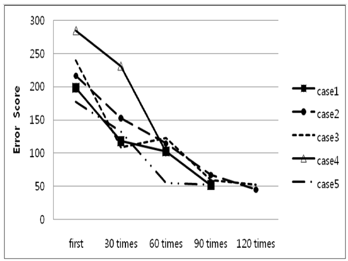
Results in Table 1 indicate that the number of patients who failed the test has decreased. After receiving 90 therapy sessions, most of the subjects passed the color vision test. Figure 2 shows the decreasing score of FM-100-hue according to the increasing frequency of treatment in some subjects. They are close to the normal score after treatment.
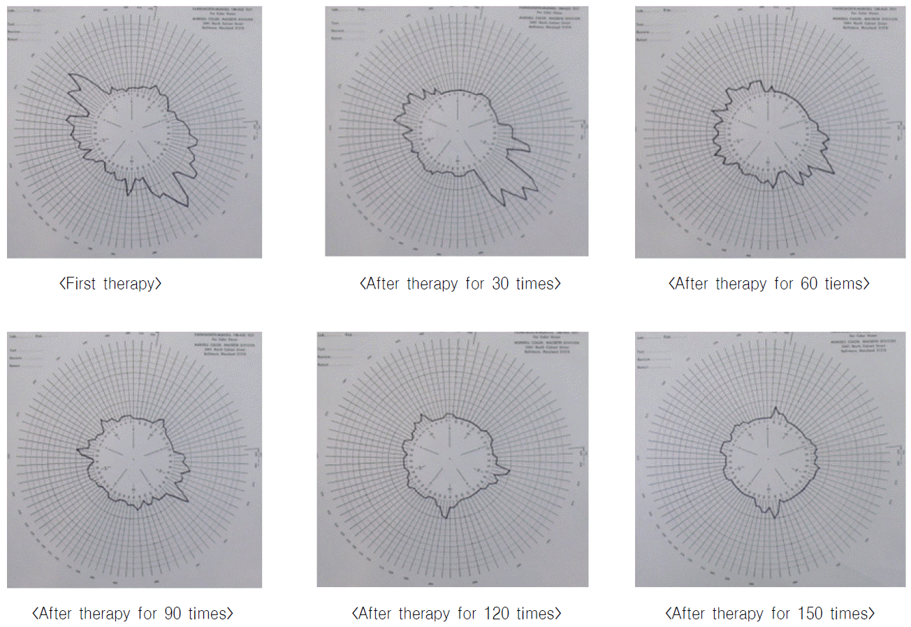
Table 3, Figures 3 and 4 show the treatment results of the representative subject. For example, a male patient, age 24, on his first hospital visit failed transformation and vanishing plates in the Ishihara test and couldn’t read 8 out of 10 screening plates in the Hahn color vision test. He was classified as deutan by both tests. According to the AO HRR test, he was classified as an R-G defect and the total errors showed a mild deutan: 7 protan and 8 deutan. He had a severe deutan defect with 12 of across the line in both test and retest by the Farnsworth D15 test. His error score was 216.5 and he was also diagnosed as having severe deuteranomalous trichromatism, including deutan axis of confusion in the F-M hue test.
After 30 sessions of therapy, he distinguished 4 out of 6 transformation plates as well as 5 out of 6 vanishing plates in the Ishihara test. He could also distinguish hidden digits and classification plates. He passed the Hahn color vision test. He could read the diagnosis series with the following error total: 9 protan and 9 deutan. However, he still made errors in the AO HRR test screening series. He passed the Farnsworth D15 test. He had a lower error score of 152.5, but still displayed the same deutan axis of confusion in the F-M 100 hue test. After 60 therapy sessions, he passed the Ishihara test, but displayed the same errors in the screening series for the AO HRR test. He had no axis of confusion with an error score of 114.5 in the F-M 100 hue test. After 90 therapy sessions, he passed the AO HRR test and had a lower error score of 67 in the F-M 100 hue test. After 120 therapy sessions, he had an even lower error score of 45 in the F-M 100 hue test. After 150 therapy sessions, he had an error score of 41 in the F-M 100 hue test, which was slightly blue-yellow. Eventually, he improved from severe deuteranomalous trichromatism to slight deuteranomalous trichromatism after 150 therapy sessions (Table 3).
The test results show the patient having normal color vision after, at least, 30 therapy sessions.
Discussion
Until present day, most doctors have not attempted to treat color vision defects by medical therapy because they had concluded that the disease is genetic and believed to be impossible to cure.
Only some doctors have tried to cure color vision defects to aid in various performances; these cures included electrical stimulation, injection of iodine, staggering doses of vitamins, flashing light therapy, and tuition by which patients were taught color naming or coached by their intuition in color1. However, patients with color vision defects are still restricted in job selection in sectors such as air and ground transportation operation, firefighting, policing, design, photography, and painting because of indeterminate and/or ineffective treatment worldwide. If patients can receive treatment, they will experience a greater quality of life. Oriental medicine approaches disease treatment with the notion that diseases originate mostly from congenital factors, which can be improved by acquired treatment. Having treated people with color vision defects, we certify the effectiveness of our treatment. The key aspect of color vision therapy was acupuncture on targeted spots to activate the retina and optic nerves relevant to eye function.
Treatment for color vision correction was used for improving the ability to perceive color by using plates corresponding to patients’ stage of color vision defects. In fact, the researchers underlined the importance of the duration of treatment and the willingness of the patient to be treated. Color vision defects cannot be guaranteed to be cured in the shortest period of time as in one of our studies. It would require patients to undergo, at least, 90 to over 120 therapy sessions, and sometimes 180+ times, to recover color vision similar to the normal seeing population.
An important part of therapy is evaluating precisely how the patient perceives his or her own state of color vision. That is, we should first distinguish between the red defect and green defect to decide the level of severity, allowing us to determine the range of perception. The range of color vision defects varies from person to person. As a matter of fact, patients with red and green defects are generally deficient in all of the blue, red, yellow, and green colors. There are individual differences in recovery proceedings after treatment. We can expect the therapy to be effective only when we know the range of color deficiency, as it takes longer to treat red-defect than green-defect. Thus, it is important to distinguish between red and green deficiencies because treatments are applied differently.
The improvement process looks like a staircase graph as opposed to a non-linear graph. In other words, improvement with treatment does not take place steadily or at regular intervals. At one point, improvement of color vision recognition comes out while it has not come out for a certain period of treatment time. The error score of the F-M 100 hue test also differs case by case. In some cases, the decrease in the score was greater during 30 therapy sessions. In a few cases, the decrease of the score was greater after a repetition of another 30 therapy sessions. In other cases, the score decreased steadily. There was a correlation to acclimatization for treatment and ability discrimination.
Through our treatment, patients often say my vision has brightened or my vision has changed beautifully during treatment. Once, a patient became extremely delighted for suddenly being able to see that his jacket was dark green and not black as he used to think, up to that moment. In conclusion, therapy to correct color vision loss is a viable option for those wishing to enjoy a better quality of life and unlimited job opportunities.


