Home | Register | Login | Inquiries | Alerts | Sitemap |  


Advanced Search
JKM > Volume 44(3); 2023 > Article
Huh and Kim: The Study on Antioxidant and Anti-inflammatory Effects of Taraxacum platycarpum H. Dahlstedt, Lonicera japonica Thunberg and Leonurus japonicus Houtt. Complex

Abstract

Objectives

This study was designed to experiment with the antioxidant and anti-inflammatory effects of Taraxacum platycarpum H. Dahlstedt, Lonicera japonica Thunberg, and Leonurus japonicus Houtt. complex (TLL) in LPS-induced RAW264.7 cell.

Methods

The antioxidant activity of TLL was measured by FRAP assay, DPPH radical scavenging activity, ABTS radical scavenging activity. Total polyphenol and flavonoid contents of TLL were measured by using standard methods. The anti-inflammatory effects of TLL were measured by NO production, biomarker production (PGE2, IL-1 β, IL-6, TNF-α), mRNA expression level (iNOS, COX-2, IL-1β, IL-6, TNF-α) and protein expression level (ERK, JNK, p38).
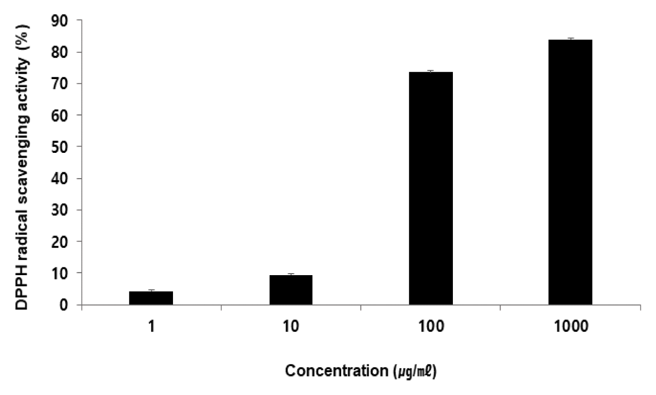
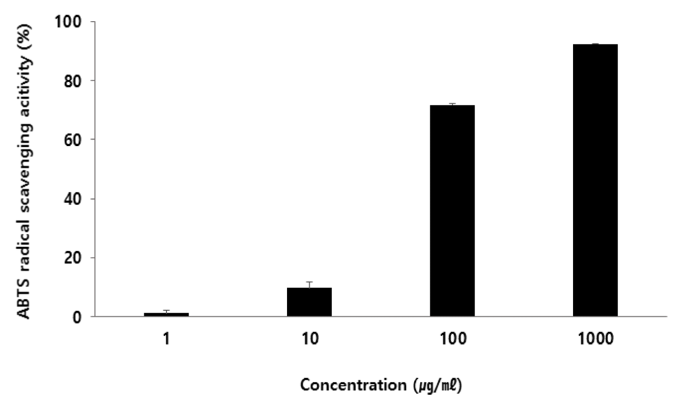
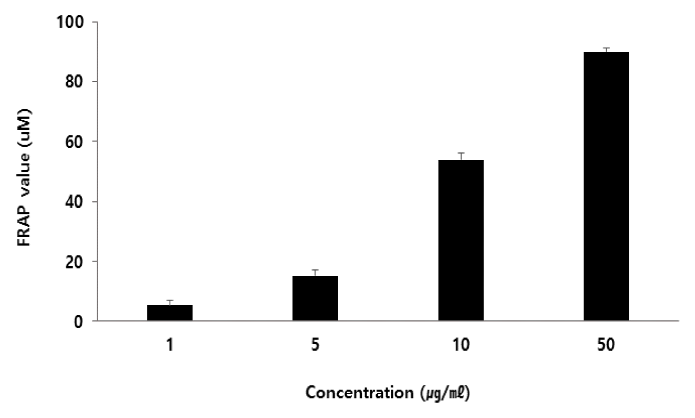
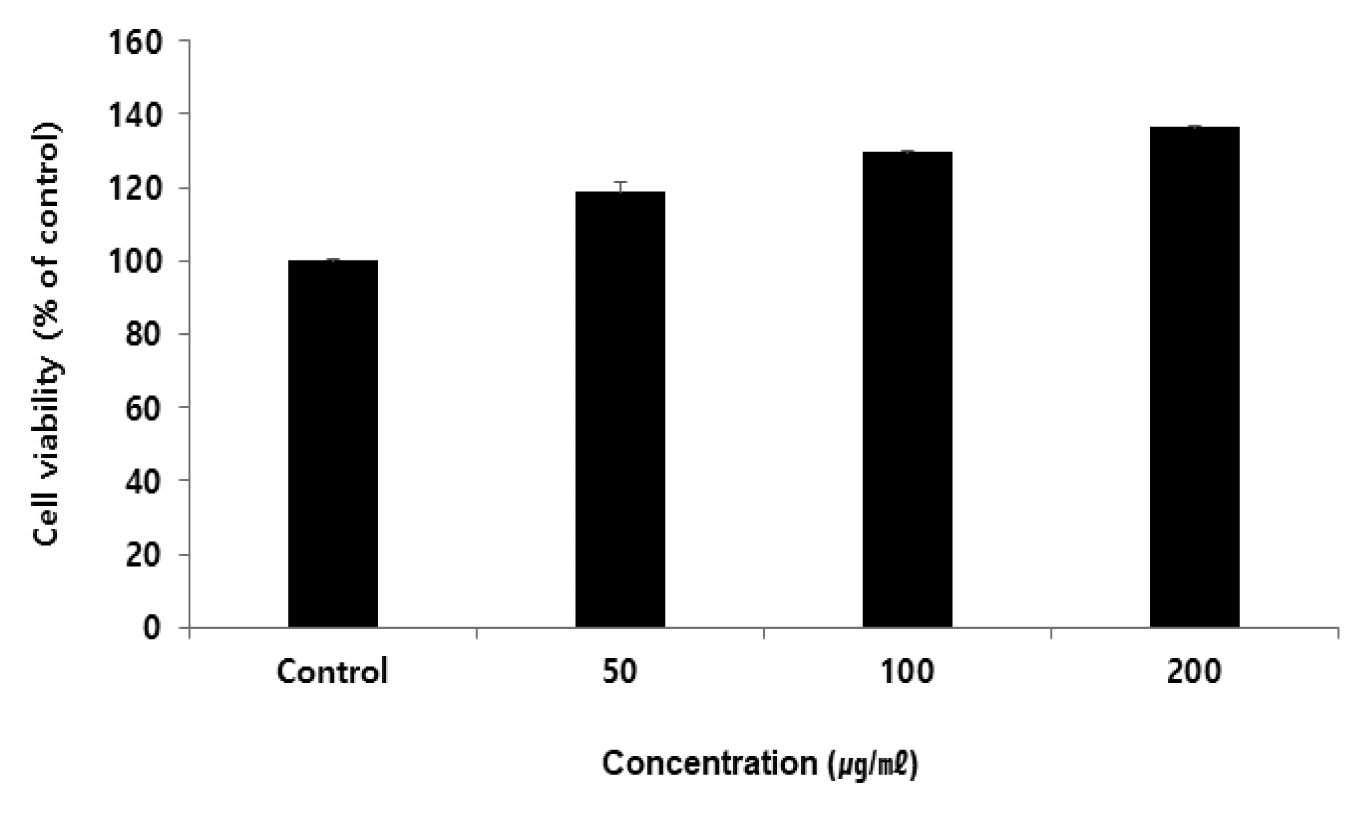
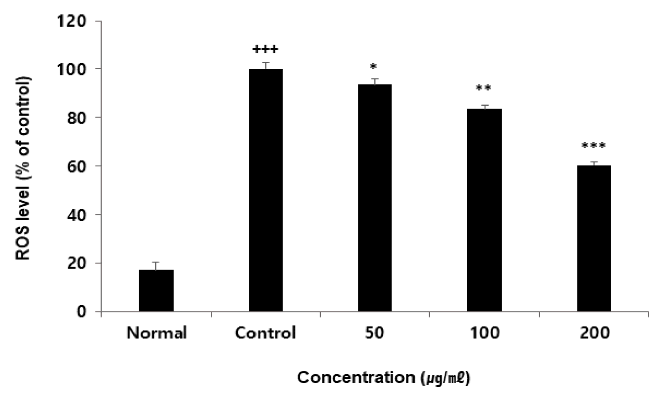
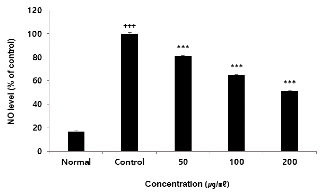
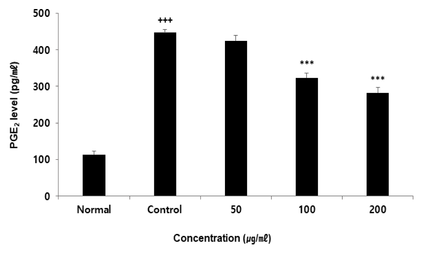
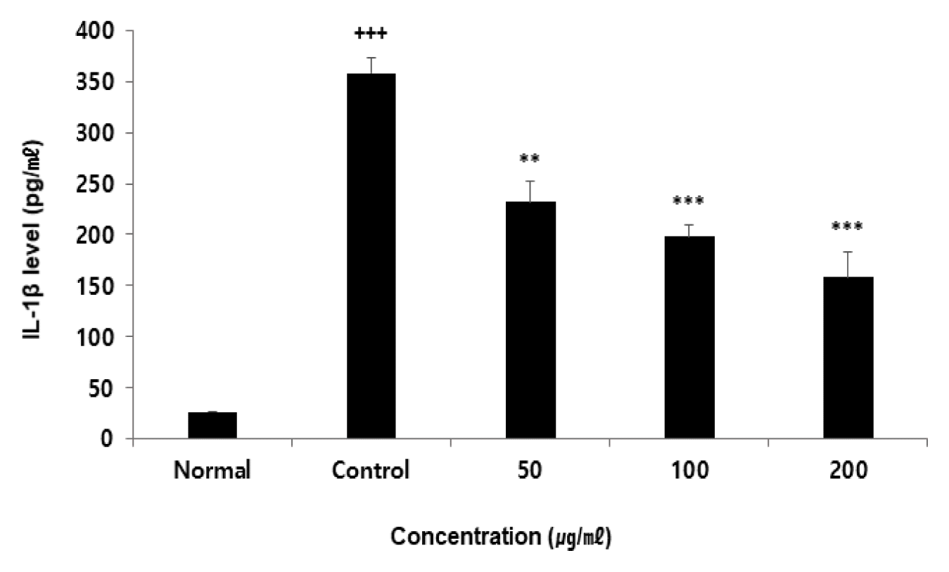
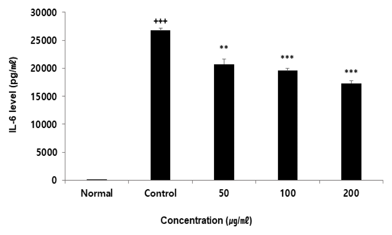
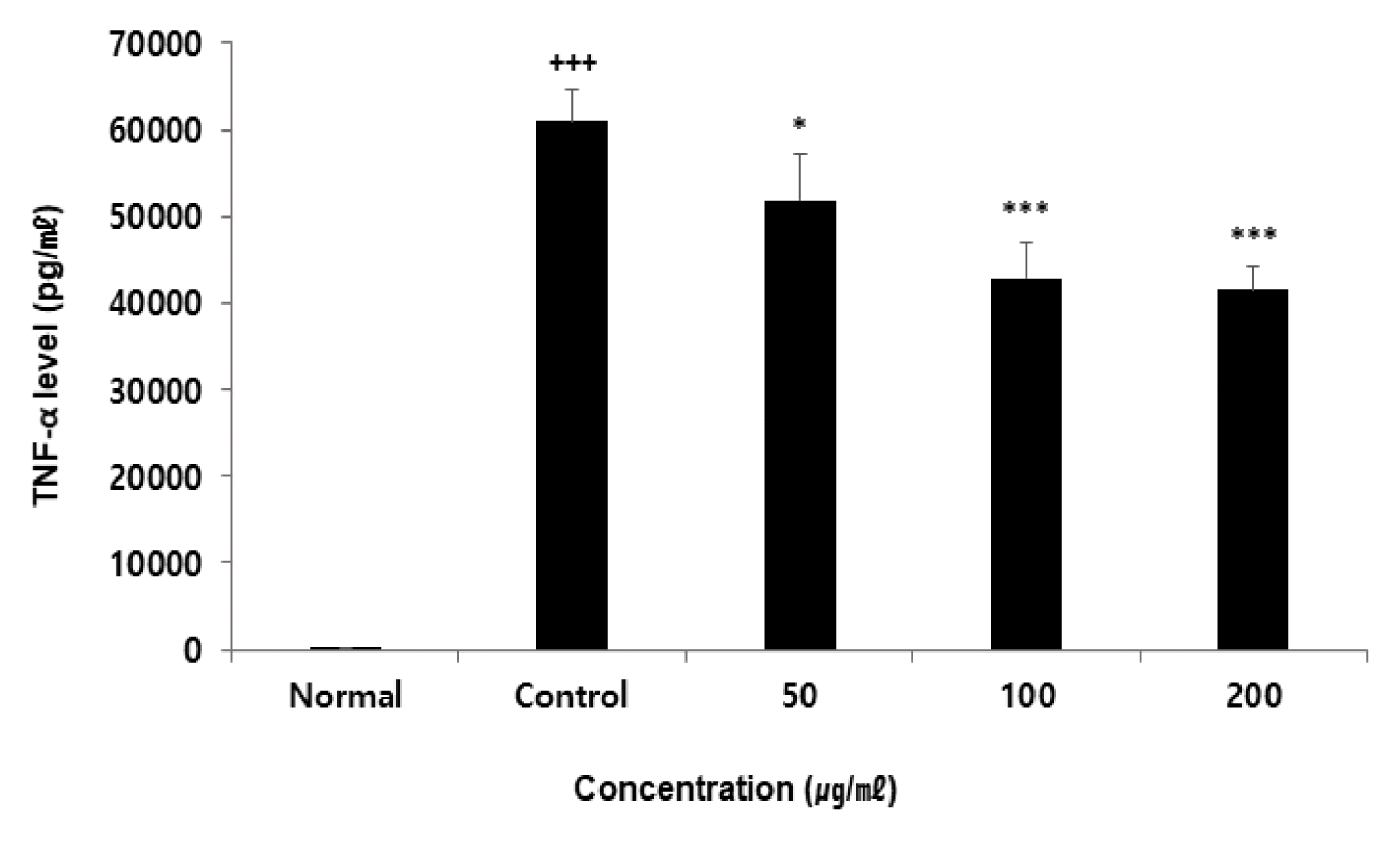
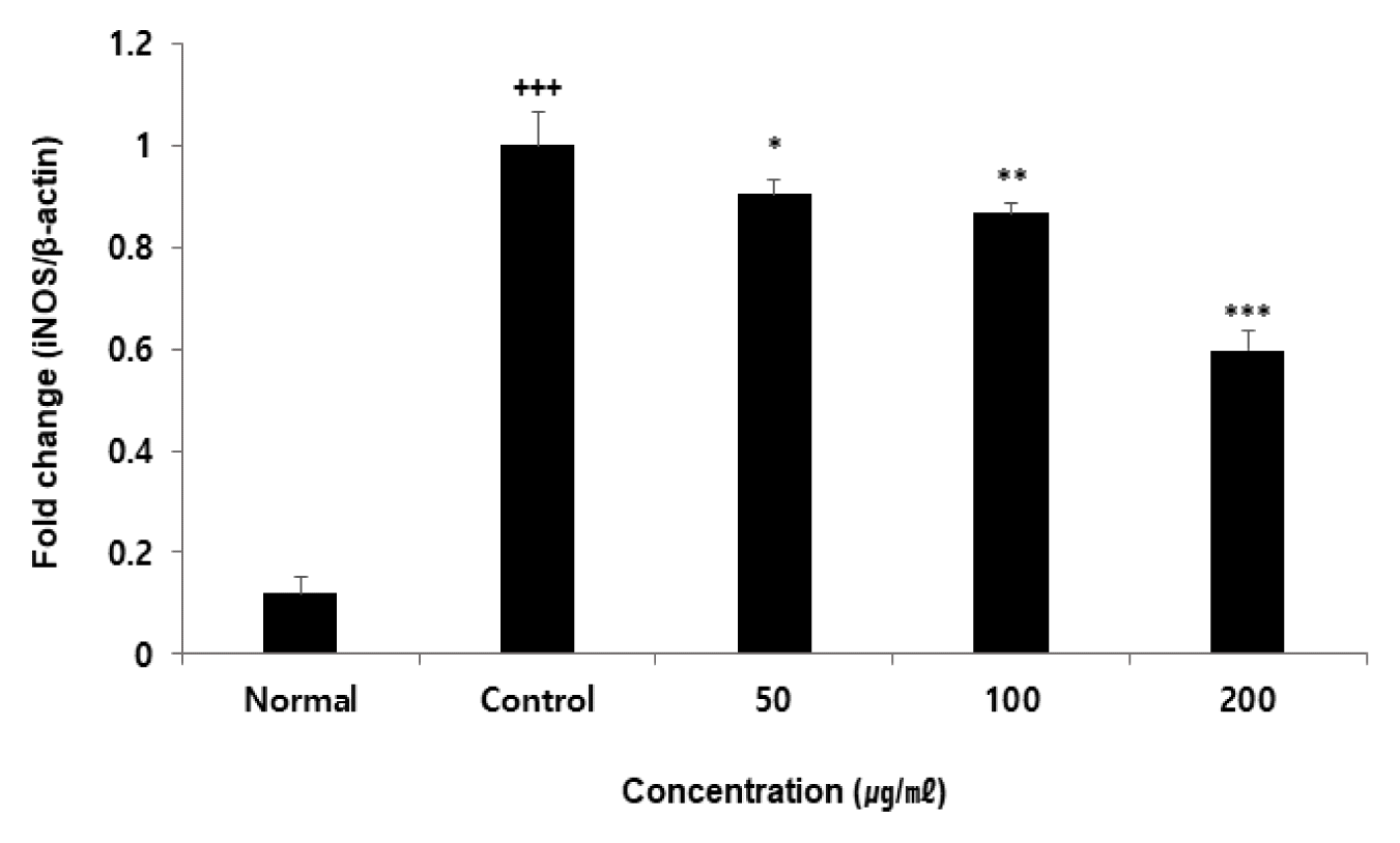
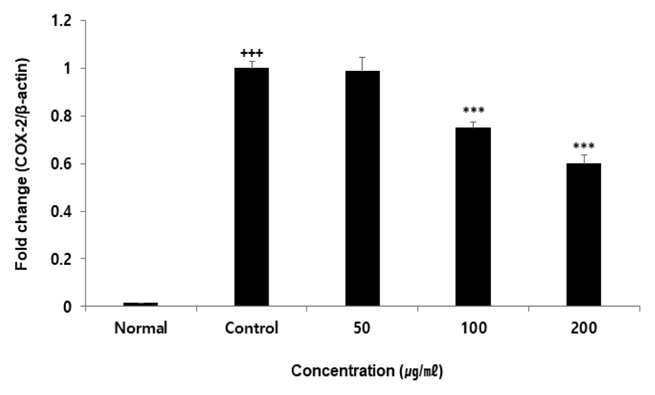
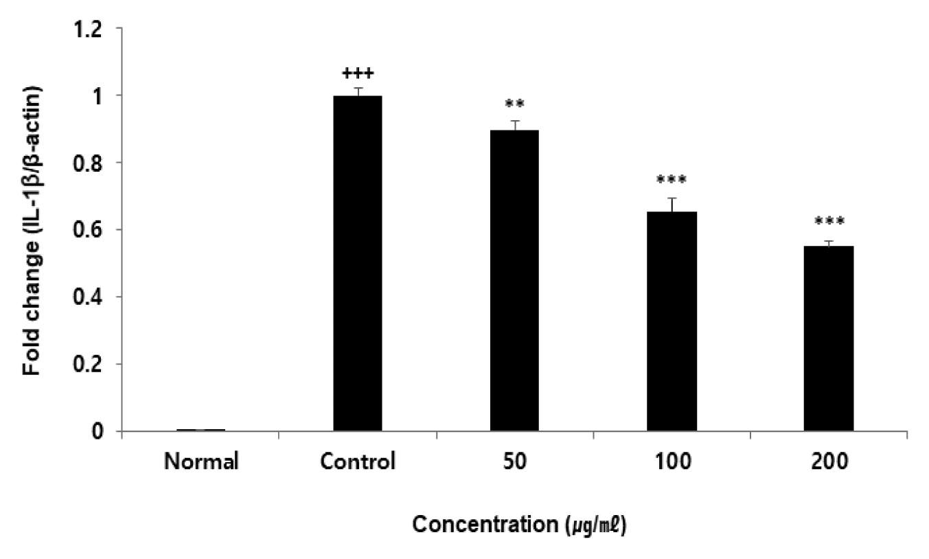
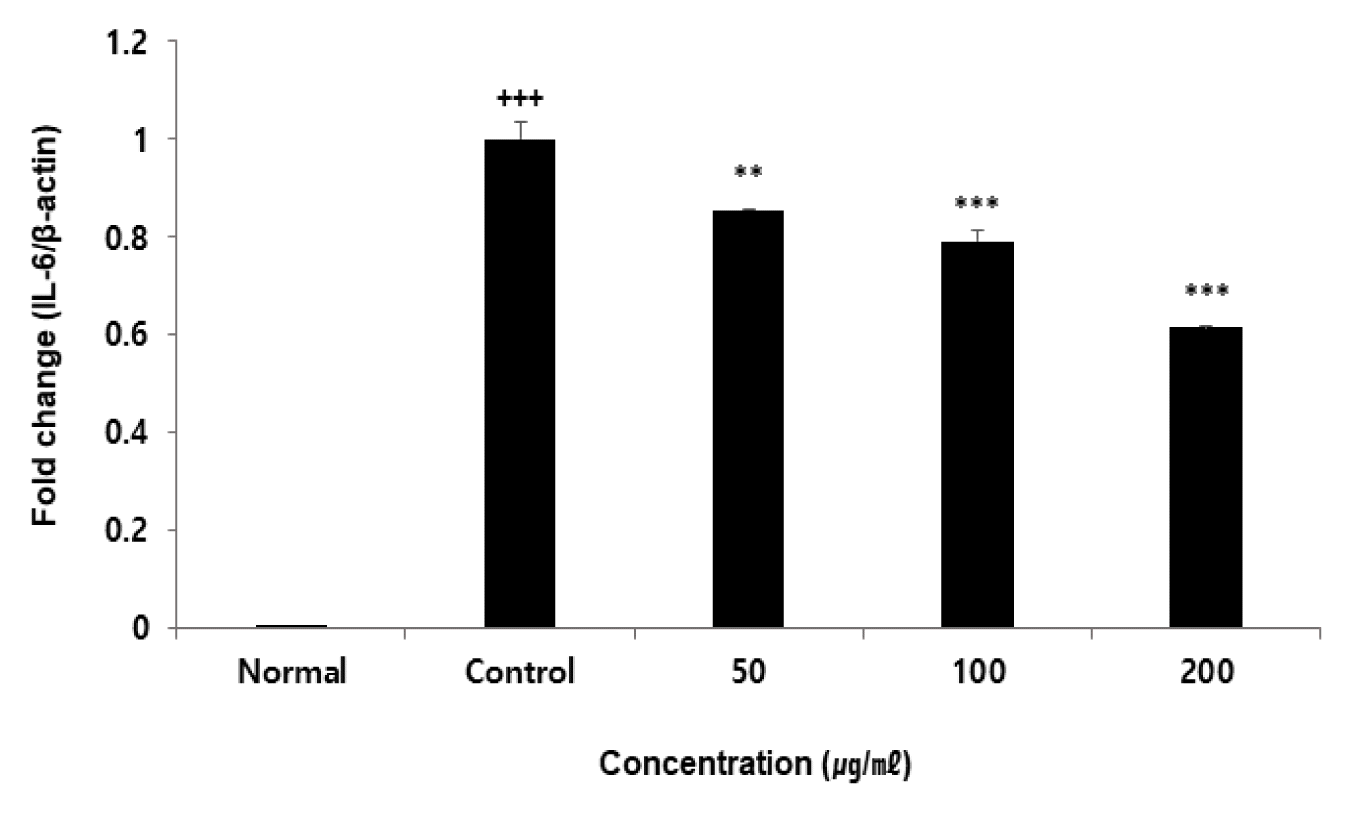
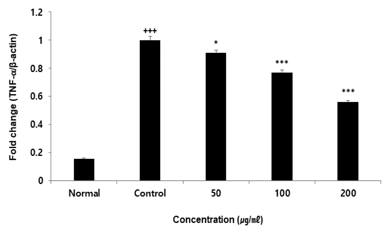
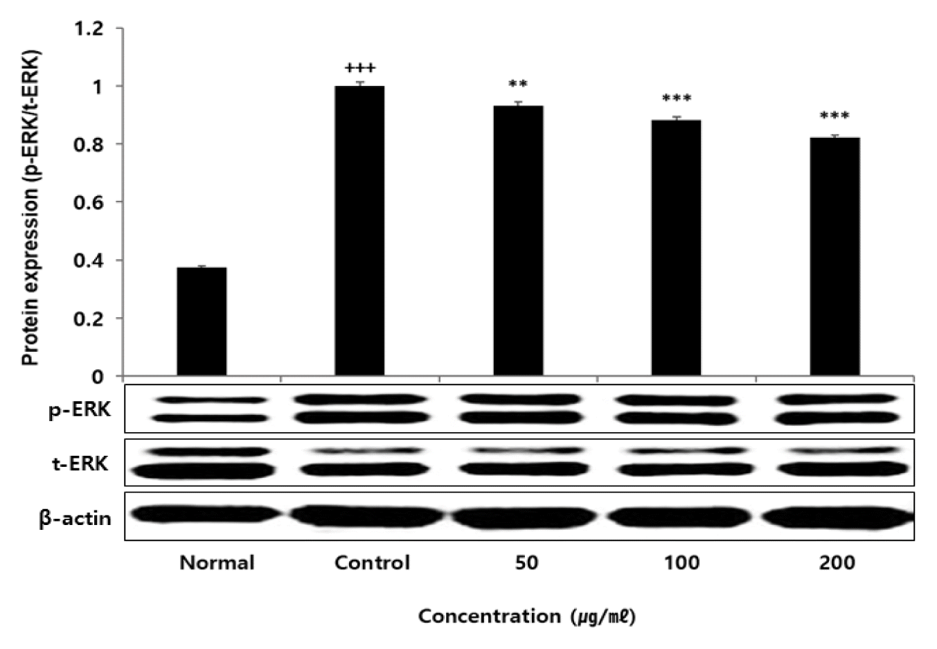
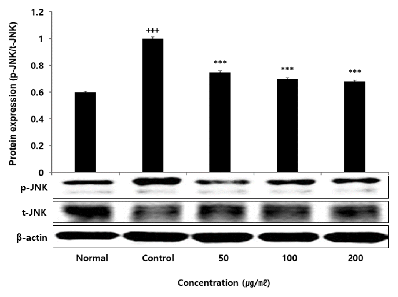
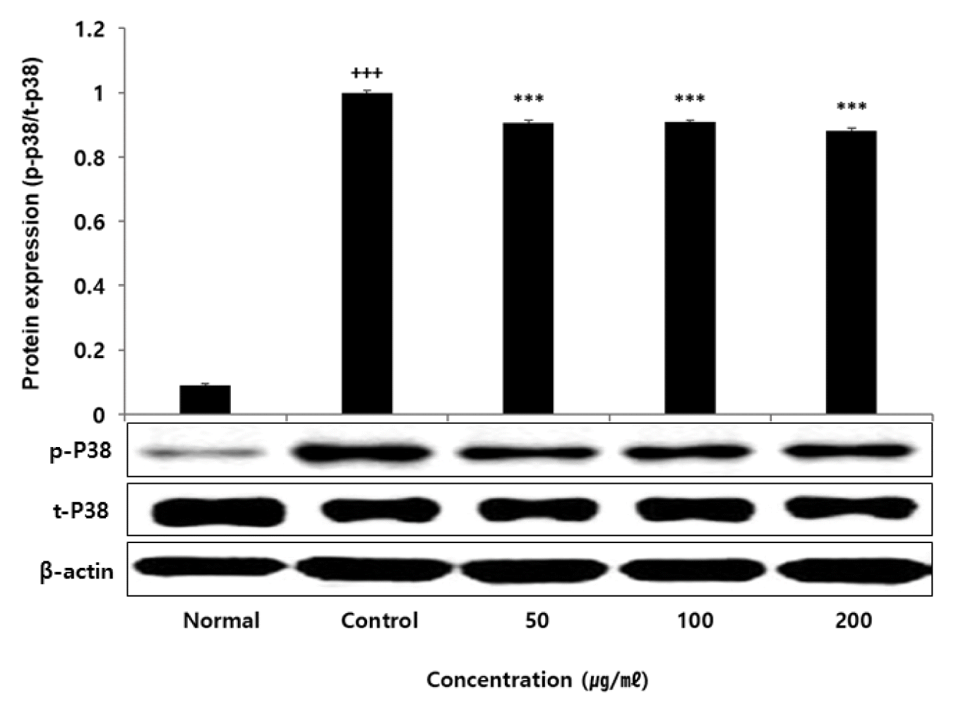
Results

Total polyphenol and flavonoid contents in TLL were 58.03±1.02 mg of Gallic acid equivalents (GAE)/g and 16.58±0.60 mg of Quercetin equivalents (QE)/g respectively. In FRAP assay, DPPH and ABTS radical scavenging activity, a concentration-dependent increase in TLL was observed. To explore antioxidant and anti-inflammatory effects of TLL, RAW 264.7 cells were treated with TLL and LPS for 24 hours. Cell viability of RAW 264.7 cells were measured by adding EZ-Cytox, It was remarkably increased at 50, 100, 200 μg/ml concentrations of TLL. NO, ROS, iNOS, IL-1β, IL-6, TNF-α, ERK, JNK and p38 were remarkably decreased at 50, 100, 200 μg/ml concentrations of TLL compared to the control group. PGE2 and COX-2 were remarkably decreased at 100, 200 μg/ml concentrations

Conclusion

These results suggest that TLL complex has antioxidant and anti-inflammatory effects.

jkm-44-3-10e1.gif
jkm-44-3-10e2.gif

Fig. 1
DPPH Radical Scavenging Activity of TLL
Each concentration (1, 10, 100, and 1000 μg/ml) of TLL was incubated with DPPH solution for 30 min. Activities were determined by measuring absorbance at 517 nm. The results were provided as mean ± SEM (n=3)
jkm-44-3-10f1.gif
Fig. 2
ABTS Radical Scavenging Activity of TLL
Each concentration (1, 10, 100, and 1000 μg/ml) of TLL was incubated with ABTS solution for 10 min. Activities were determined by measuring absorbance at 732 nm. The results were provided as mean ± SEM (n=3)
jkm-44-3-10f2.gif
Fig. 3
FRAP Value of TLL.
Each concentration (1, 5, 10, and 50 μg/ml) of TLL was incubated with FRAP regent solution for 30 min. Activities were determined by measuring absorbance at 593 nm. The results were provided as mean ± SEM (n=3)
jkm-44-3-10f3.gif
Fig. 4
Cell viability of TLL in RAW264.7 cell.
RAW264.7 cells were treated by each concentration (50, 100, and 200 μg/ml) of TLL for 24 h. EZ-Cytox was added to treated cells and reacted for 30 min, thereafter microplate reader was used for measuring absorbance of the solution at 450 nm. Cell viability was calculated as percentage with regard to the control. The results were provided as mean ± SEM (n=3)
jkm-44-3-10f4.gif
Fig. 5
Effect of TLL on ROS level in RAW264.7 cell
RAW264.7 cells were treated by each concentration (50, 100 and 200 μg/ml) of TLL with 100 ng/ml LPS for 24 h. ROS levels were calculated as percentage with regard to the control. The results were provided as mean ± SEM (n=3) (Significance of results, +++: p<0.001 compared to normal, *: p<0.05, **: p<0.01, ***: p <0.001 compared to control).
jkm-44-3-10f5.gif
Fig. 6
Effect of TLL on NO level in RAW264.7 cell
RAW264.7 cells were treated by each concentration (50, 100 and 200 μg/ml) of TLL with 100 ng/ml LPS for 24 h. NO levels were calculated as percentage with regard to the control. The results were provided as mean ± SEM (n=3) (Significance of results, +++: p<0.001 compared to normal, ***: p<0.001 compared to control).
jkm-44-3-10f6.gif
Fig. 7
Effect of TLL on PGE2 level in RAW264.7 cell
RAW264.7 cells were treated by each concentration (50, 100, and 200 μg/ml) of TLL with 100 ng/ml LPS for 24 h. Treated cells were measured by mouse PGE2 ELISA kit. The results were provided as mean ± SEM (n=3) (Significance of results, +++: p<0.001 compared to normal, ***: p<0.001 compared to control).
jkm-44-3-10f7.gif
Fig. 8
Effect of TLL on IL-1β level in RAW264.7 cell
RAW264.7 cells were treated by each concentration (50, 100, and 200 μg/ml) of TLL with 100 ng/ml LPS for 24 h. Treated cells were measured by mouse IL-1beta ELISA kit. The results were provided as mean ± SEM (n=3) (Significance of results, +++: p<0.001 compared to normal, **: p<0.01, ***: p<0.001 compared to control).
jkm-44-3-10f8.gif
Fig. 9
Effect of TLL on IL-6 level in RAW264.7 cell
RAW264.7 cells were treated by each concentration (50, 100, and 200 μg/ml) of TLL with 100 ng/ml LPS for 24 h. Treated cells were measured by mouse IL-6 ELISA kit. The results were provided as mean ± SEM (n=3) (Significance of results, +++: p<0.001 compared to normal, **: p<0.01, ***: p<0.001 compared to control).
jkm-44-3-10f9.gif
Fig. 10
Effect of TLL on TNF-α level in RAW264.7 cell
RAW264.7 cells were treated by each concentration (50, 100, and 200 μg/ml) of TLL with 100 ng/ml LPS for 24 h. Treated cells were measured by mouse TNF-α ELISA kit. The results were provided as mean ± SEM (n=3) (Significance of results, +++: p<0.001 compared to normal, *: p<0.05, ***: p<0.001 compared to control).
jkm-44-3-10f10.gif
Fig. 11
Effect of TLL on iNOS mRNA expression level in RAW264.7 cell
RAW264.7 cells were treated by each concentration (50, 100, and 200 μg/ml of TLL with 100 ng/ml) LPS for 24 h. The mRNA levels of iNOS expression were measured by polymerase chain reaction. The normal group was not treated LPS. The results were provided as mean ± SEM (n=3) (Significance of results, +++: p<0.001 compared to normal, *: p<0.05, **: p<0.01, ***: p<0.001 compared to control).
jkm-44-3-10f11.gif
Fig. 12
Effect of TLL on COX-2 mRNA expression level in RAW264.7 cell
RAW264.7 cells were treated by each concentration (50, 100, and 200 μg/ml) of TLL with 100 ng/ml LPS for 24 h. The mRNA levels of COX-2 expression were measured by polymerase chain reaction. The normal group was not treated LPS. The results were provided as mean ± SEM (n=3) (Significance of results, +++: p<0.001 compared to normal, ***: p<0.001 compared to control).
jkm-44-3-10f12.gif
Fig. 13
Effect of TLL on IL-1β mRNA expression level in RAW264.7 cell
RAW264.7 cells were treated by each concentration (50, 100, and 200 μg/ml) of TLL with 100 ng/ml LPS for 24 h. The mRNA levels of IL-1β expression were measured by polymerase chain reaction. The normal group was not treated LPS. The results were provided as mean ± SEM (n=3) (Significance of results, +++: p<0.001 compared to normal, **: p<0.01, ***: p<0.001 compared to control).
jkm-44-3-10f13.gif
Fig. 14
Effect of TLL on IL-6 mRNA expression level in RAW264.7 cell
RAW264.7 cells were treated by each concentration (50, 100, and 200 μg/ml) of TLL with 100 ng/ml LPS for 24 h. The mRNA levels of IL-6 expression were measured by polymerase chain reaction. The normal group was not treated LPS. The results were provided as mean ± SEM (n=3) (Significance of results, +++: p<0.001 compared to normal, **: p<0.01, ***: p<0.001 compared to control).
jkm-44-3-10f14.gif
Fig. 15
Effect of TLL on TNF-α mRNA expression level in RAW264.7 cell
RAW264.7 cells were treated by each concentration (50, 100, and 200 μg/ml) of TLL with 100 ng/ml LPS for 24 h. The mRNA levels of TNF-α expression were measured by polymerase chain reaction. The normal group was not treated LPS. The results were provided as mean ± SEM (n=3) (Significance of results, +++: p <0.001 compared to normal, *: p<0.05, ***: p<0.001 compared to control).
jkm-44-3-10f15.gif
Fig. 16
Effect of TLL on ERK protein expression level in RAW264.7 cell
RAW264.7 cells were treated by each concentration (50, 100, and 200 μg/ml) of TLL with 100 ng/ml LPS for 24 h. The total cell extracts were subjected to western blot and 10% SDS-PAGE analysis with the respective primary and secondary antibodies. The results were provided as mean ± SEM (n=3) (Significance of results, +++: p<0.001 compared to normal, **: p<0.01, ***: p <0.001 compared to control).
jkm-44-3-10f16.gif
Fig. 17
Effect of TLL on JNK protein expression level in RAW264.7 cell
RAW264.7 cells were treated by each concentration (50, 100, and 200 μg/ml) of TLL with 100 ng/ml LPS for 24 h. The total cell extracts were subjected to western blot and 10% SDS-PAGE analysis with the respective primary and secondary antibodies. The results were provided as mean ± SEM (n=3) (Significance of results, +++: p<0.001 compared to normal, ***: p<0.001 compared to control).
jkm-44-3-10f17.gif
Fig. 18
Effect of TLL on p38 protein expression level in RAW264.7 cell
RAW264.7 cells were treated by each concentration (50, 100, and 200 μg/ml) of TLL with 100 ng/ml LPS for 24 h. The total cell extracts were subjected to western blot and 10% SDS-PAGE analysis with the respective primary and secondary antibodies. The results were provided as mean ± SEM (n=3) (Significance of results, +++: p<0.001 compared to normal, ***: p<0.001 compared to control).
jkm-44-3-10f18.gif
Table 1
The Sequence of Primers in Real-Time PCR
Primer Forward / Reverse Sequence (5'→3')
iNOS Forward CGAAACGCTTCACTTCCAA
Reverse TGAGCCTATATTGCTGTGGCT

IL-1β Forward AAGAAGAGCCCATCCTCTGT
Reverse GGAGCCTGTAGTGCACTTGT

IL-6 Forward AGTCCTTCCTACCCCAATTTCC
Reverse GGTCTTGGTCCTTAGCCACT

TNF-α Forward ATGGCCTCCCTCTCATCAGT
Reverse TTTGCTACGACGTGGGCTAC

COX-2 Forward AACCGCATTGCCTCTGAAT
Reverse CATGTTCCAGGAGGATGGAG

β-actin Forward AGGGAAATCGTGCGTGACAT
Reverse TCCAGGGAGGAAGAGGATGC

참고문헌

1. Reece JB, Urry LA, Cain ML, Wasserman SA, Minorsky PV, Jackson R.BCampbell Biology. 10th ed. Seoul: Bio Science;2016. p. 202–6.


2. Pack JH, Kweon GR. 2013; Clinical Applications of Antioxidants. Hanyang Medical Reviews. 33:2. 130–6. http://dx.doi.org/10.7599/hmr.2013.33.2.130
crossref

3. Coyle JT, Puttfarcken P. 1993; Oxidative stress, glutamate, and neurodegenerative disorders. Science. 262:5134. 689–95. 10.1126/science.7901908
crossref pmid

4. Kwag SG, Kwon SY, Kim SK, Min BW, Seo YM, Lee JM, et al. Basic Pathology. Seoul: Jungmunkag;2010. p. 84


5. Punt J, Stranford SA, Jones PP, Owen JA. Kuby Immunology. 8th ed. Seoul: Panmunedu;2020. p. 142–3.


6. Abbas AK, Lichtman AH, Pillai S. Basic Immunology: Functions and Disoders of the Immune System. 6th ed. Seoul: Panmunedu;2020. p. 35p. 44p. 47


7. Park MG, Yoo JD, Lee KH. 2020; Current Guidelines for Non-Steroidal Anti-Inflammatory Drugs. J Korean Orthop Assoc. 55:1. 9–28. https://doi.org/10.4055/jkoa.2020.55.1.9


8. Golan DE, Armstrong EJ, Armstrong AW. Principles of Pharmacology: The pathophysiologic Basis of Drug Therapy. 4th ed. Seoul: Shinilbooks;2017. p. 795–8. p. 811


9. Cha HY, Jeong AR, Cheon JH, Ahn SH, Park SY, Kim KB. 2015; The Anti-oxidative and Anti-inflammatory Effect of Lonicera Japonica on Ulcerative Colitis Induced by Dextran Sulfate Sodium in Mice. Korean J Pediatr. 29:3. 54–64. http://dx.doi.org/10.7778/jpkm.2015.29.3.054
crossref

10. Choi YN, Choi YK, Nan L, Choo BK. 2020; Anti-oxidant and Anti-inflammatory Effects of Ethanol Extracts from Leonurus japonicus Houtt. on LPS-induced RAW 264.7 Cells. Korean J Org Agri. 28:4. 659–77. http://dx.doi.org/10.11625/KJOA.2020.28.4.659


11. Lee YS. 2020; Anti-aging, Antioxidant and Anti-inflammation Activities of Lonicerae Flos, Dictamni Radicis Cortex and Forsythiae Fructus Extracts. Journal of Investigative Cosmetology. 16:1. 1–10. https://doi.org/10.15810/jic.2020.16.1.001


12. Herbology Editorial Committee of Korean Medicine schools. Herbology [Boncho-hak]. 4th ed. Seoul: Young-Lim Press;2020. p. 236–8. p. 240–2. p. 452–3.


13. Noh KH, Baek JH. 2009; Inhibitory Effect of Taraxaci Herba Extract (THE) on Pro-inflammatory Mediatory. The Journal of Korean Oriental Pediatrics. 23:3. 165–76.


14. Kang JR. 2009. Anti-oxidation and Melanin Inhibition Characteristics of Taraxacum platycarpum and Compositae plants. Ph.D. thesis. Konkuk University.


15. So MS. 2009; Effect of Taraxacum mongolieum Hand-Mass on Proliferation of Breast Cancer Cells and Nitric Oxide Production. Journal of Korean Health & Fundamental Medical Science. 2:3. 128–31.


16. Jo JH, Lyu JH, Kim CH, Kang KH, Yoon HJ, Lee SY, et al. 2007; The Anti-allergic Effects of Taraxaci Herba on the RBL-2H3 Cells. J Korean Med Ophthalmol Otolaryngol Dermatol. 20:1. 209–17.


17. Lee HW, Ma CJ. 2021; Neuroprotective Activity of Lonicerin Isolated from Lonicera japonica. Kor J Pharmacogn. 52:1. 19–25. https://doi.org/10.22889/KJP.2021.52.1.19


18. Ahn SH, Kim HH. 2010; Lonicerae Flos Inhibited COX-2 and MMP-9 in LPS Induced Arthritis of Mouse through Regulation of MIF. Korean J Oriental Physiology & Pathology. 24:2. 242–8.


19. Park CW. Herbal Pharmacology [Hanyak-yakri-hak]. Seoul: Shin Kwang Pub;2005. p. 169p. 243–4.


20. Nho JH, Lee HJ, Jang JH, Yang BD, Woo KW, Kim HA, et al. 2019; Protective Effect of Water Extract of Leonurus japonicus Houttuyn against HCl/EtOH-induced Gastric Mucosal Damage and Genotoxicity Evaluation using Micronucleus Test. Korean J. Plant Res. 32:4. 282–9. https://doi.org/10.7732/kjpr.2019.32.4.282


21. Xie M. Formulas of Chinese Medicine [fangji-xue]. Beijing: People’s Medical Pubishing House;2008. p. 453–4.


22. Park JH, Park JY, Park SD. 2014; Anti-Oxidative and Anti-Inflammatory Effect of 7 Herbal Extracts and Methods of Herbal Formula Compositioning. Herbal Formula Science. 22:2. 87–103. 10.14374/HFS.2014.22.2.087
crossref

23. Hong SJ, Choi EG, Lim HS, Shon JB, Jeong SS. 2001; Effect of herbal dentifrice on dental plaque and gingivitis. Korean Academy of Dental Health. 25:4. 347–355.


24. Choi KM, Jeon JH, Kim ES, Sung KJ, Kim YI. 2021; Study on Inflammatory-Control Mechanisms of Lonicera japonica Thunberg, Forsythia viridissima Lindley, and Taraxacum platycarpum H. Dahlstedt Complex. Journal of Haehwa Medicine. 30:1. 11–31.


25. Lee SB. 2021. Anti-inflammatory Effects of Taraxacum platycarpum H. Dahlstedt, Lonicera japonica Thunberg, and Houttuynia cordata Thunberg Complex in Lipopolysaccharide-Stimulated Macrophage and Mice. M.S. thesis. Daejeon University.


26. Moon SC. 2022. Antioxidant and Anti-inflammatory Effects of Taraxacum platycarpum H. Dahlstedt, Lonicera japonica Thunberg, Citrus unshiu Markovich complex. M.S. thesis. Daejeon University.


27. Zorov DB, Juhaszova M, Sollott SJ. 2014; Mitochondrial reactive oxygen species (ROS) and ROS-induced ROS release. Physiol Rev. 94:3. 909–50. 10.1152/physrev.00026.2013
crossref pmid pmc

28. Li R, Jia Z, Trush MA. 2016; Defining ROS in Biology and Medicine. Reactive Oxygen Species (Apex). 1:1. 9–21. 10.20455/ros.2016.803
crossref pmid pmc

29. Frankel EN. 1996; Antioxidants in lipid foods and their impact on quality. Food Chemistry. 57:1. 51–5. https://doi.org/10.1016/0308-8146(96)00067-2
crossref

30. Kim HC. Herbal Pharmacology [Hanyak-yakri-hak]. Seoul: Jipmoon;2001. p. 150–2. p. 155–6. p. 334–6.


31. Yang JH, Yoo HR, Kim YS, Seol IC. 2021; The Effect of Lonicera Japonica Thunberg on Inflammatory Factor Expression Associated with Atherosclerosis. J Int Korean Med. 42:1. 25–39. https://doi.org/10.22246/jikm.2021.42.1.25


32. Kim TY, Jang SA, Chae YB, Bak JP. 2016; Antioxidant and Protective Effects of Leonurus sibiricus L. Extract on Ultraviolet B (UVB)-induced Damage in Human Keratinocytes. Korean J. Plant Res. 29:1. 11–9. http://dx.doi.org/10.7732/kjpr.2016.29.1.011


33. Yoon HJ, Moon ME, Park HS, Im SY, Kim YH. 2007; hitosan oligosaccharide (COS) inhibits LPS-induced inflammatory effects in RAW 264.7 macrophage cells. Biochemical and Biophysical Research Communications. 358:3. 954–9. 10.1016/j.bbrc.2007.05.042
crossref pmid

34. Shin JH, Yoo SK. 2012; Antioxidant Properties in Microbial Fermentation Products of Lonicera japonica Thunb. Extract. Journal of the East Asian Society of Dietary Life. 22:1. 95–102.


35. Ozben T. 2007; Oxidative stress and apoptosis: impact on cancer therapy. Journal of Pharmaceutical Sciences. 96:9. 2181–96.
crossref pmid

36. Moloney JN, Cotter TG. 2018; ROS signalling in the biology of cancer. Semin Cell Dev Biol. 80:50–64. 10.1016/j.semcdb.2017.05.023
crossref pmid

37. Yim CY. 2010; Nitric oxide and cancer. Korean J Med. 78:4. 430–6.


38. Jin JR, Kim JH, Lim JH, Jung YH, Kim KH. PP+ Immunology. Seoul: Life Science;2020. p. 40


39. Kim MJ, Park SH, Kim KBWR, Choi JS, Ahn DH. 2017; Anti-Inflammatory Effect of Chondria crassicaulis Ethanol Extract on MAPKs and NF-κB Signaling Pathway in LPS-Induced RAW 264.7 Macrophages. KSBB Journal. 32:4. 352–60. http://dx.doi.org/10.7841/ksbbj.2017.32.4.352
crossref

40. Chan ED, Riches DW. 2001; IFN-gamma + LPS induction of iNOS is modulated by ERK, JNK/SAPK, and p38(mapk) in a mouse macrophage cell line. Am J Physiol Cell Physiol. 280:3. C441–50. https://doi.org/10.1152/ajpcell.2001.280.3.C441
pmid

41. Murakami-Mori K, Mori S, Nakamura S. 1999; p38MAP kinase is a negative regulator for ERK1/2-mediated growth of AIDS-associated Kaposi's sarcoma cells. Biochem Biophys Res Commun. 264:3. 676–82. https://doi.org/10.1006/bbrc.1999.1574
crossref pmid

TOOLS
PDF Links  PDF Links
Full text via DOI  Full text via DOI
PubReader  PubReader
Download Citation  Download Citation
  Print
Share:      
METRICS
0
Crossref
1,135
View
51
Download
Editorial office contact information
3F, #26-27 Gayang-dong, Gangseo-gu Seoul, 157-200 Seoul, Korea
The Society of Korean Medicine
Tel : +82-2-2658-3627   Fax : +82-2-2658-3631   E-mail : skom1953.journal@gmail.com
About |  Browse Articles |  Current Issue |  For Authors and Reviewers
Developed in M2PI