Home | Register | Login | Inquiries | Alerts | Sitemap |  


Advanced Search
JKM > Volume 45(1); 2024 > Article
Jeon, Lee, Kwon, Cho, Jung, Moon, Park, Ko, and Park: The Analysis of relation to brain iron deposition of Parkinson’s Disease using Quantitative Susceptibility Mapping

Abstract

Objectives

This study aimed to investigate the levels of brain iron deposition in Parkinson’s disease (PD) patients using Quantitative Susceptibility Mapping (QSM) and to determine whether distinctions compared to the general population exist. Furthermore, we examined potential variations in iron deposition among different PD subtypes.
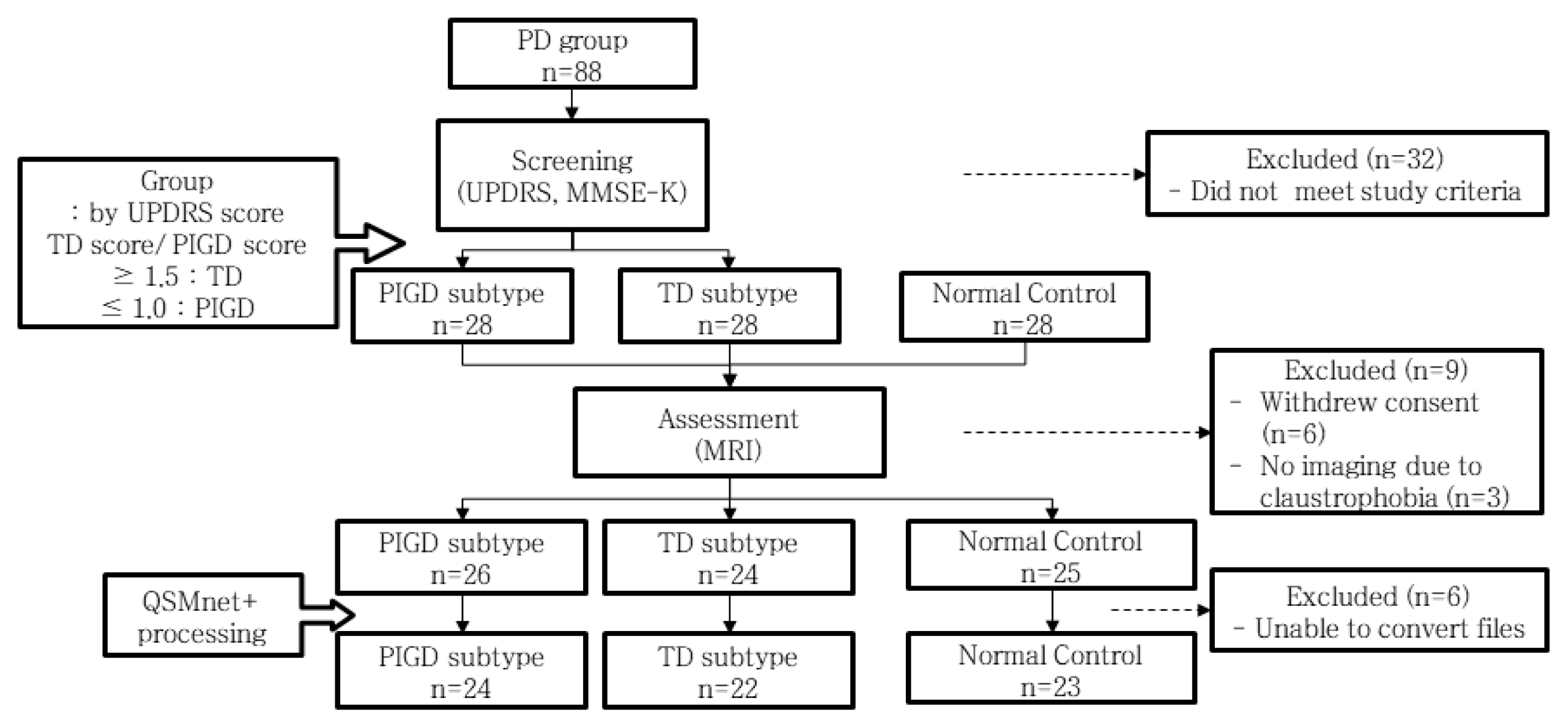
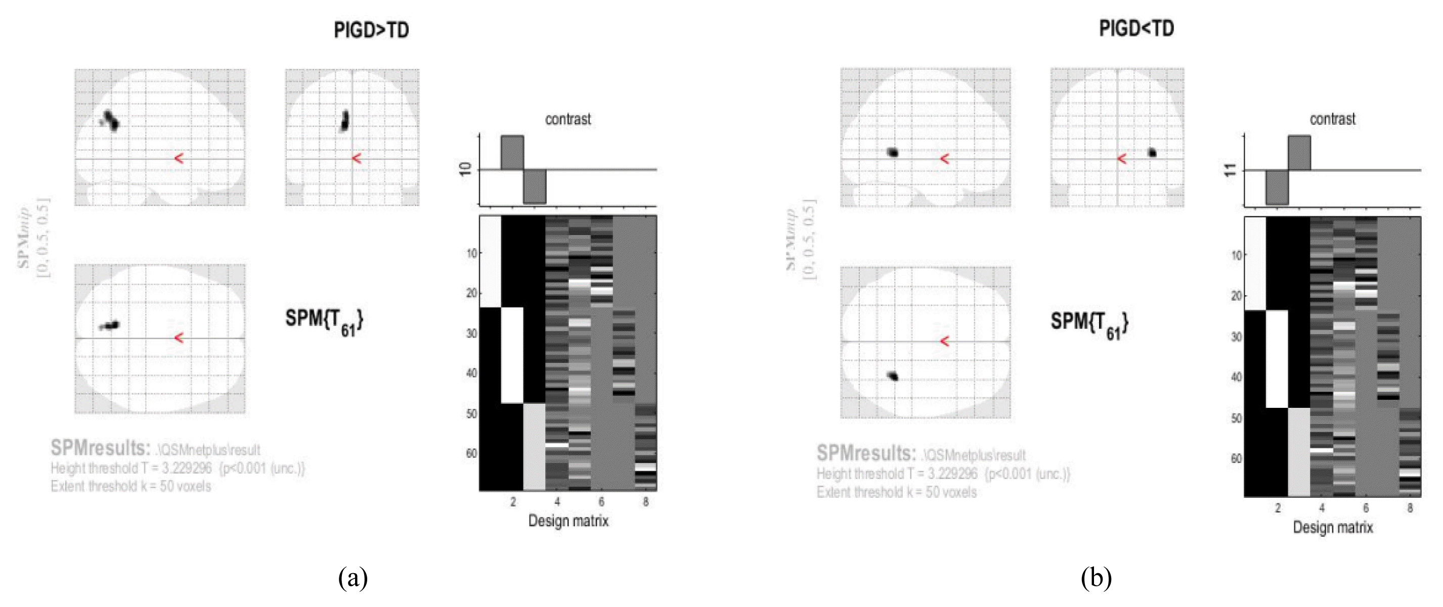
Methods

Structural brain imaging was conducted on 75 participants at Gangdong Kyung Hee University Hospital between August 2017 and May 2020. PD patients were categorized into Tremor Dominant (TD) and Postural Instability and Gait Difficulty (PIGD) subtypes. Voxel-based morphometry and QSM were employed to compare voxel-wise magnetic susceptibility across the entire brain between Normal Controls (NC) and PD groups. Subsequently, QSM values were compared between TD and PIGD groups.
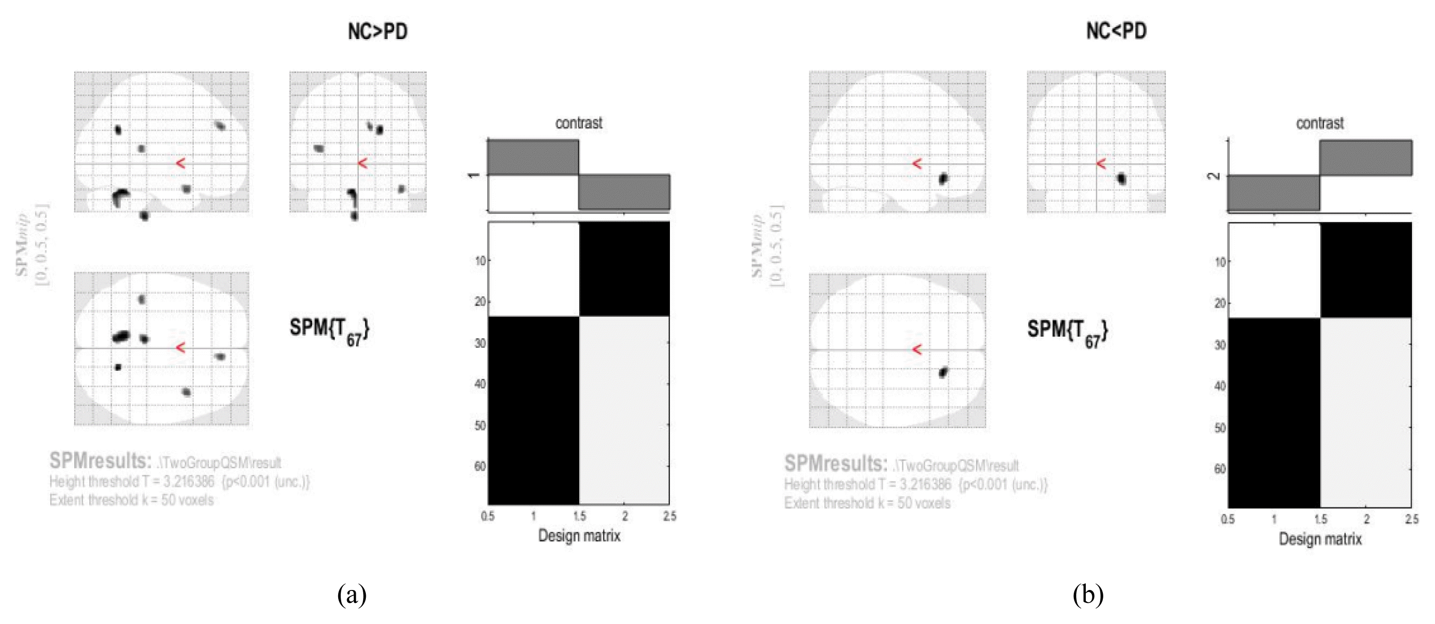
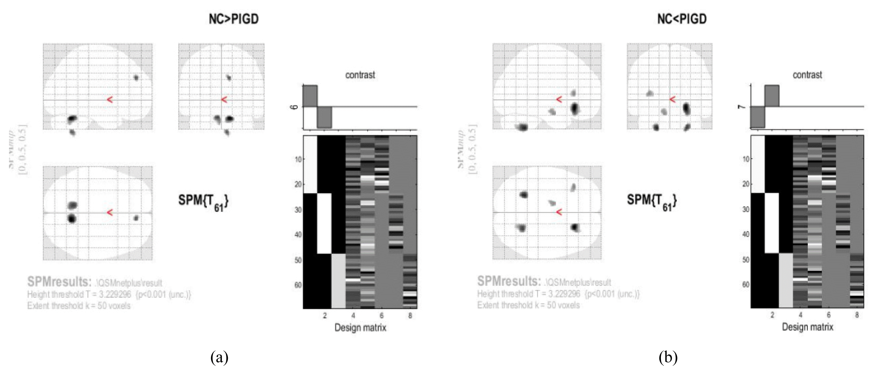
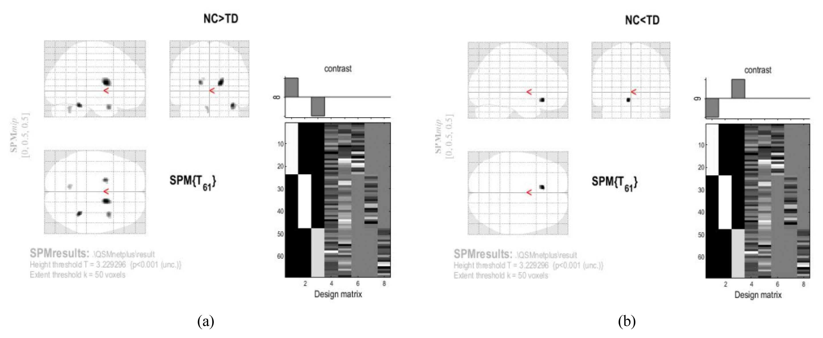
Results

QSM values were compared among 46 PD patients and 23 normal controls, as well as between TD (n=22) and PIGD (n=24) groups. Voxel-based QSM analysis revealed no significant differences between groups. Similarly, ROI-based QSM analysis showed no significant distinctions.

Conclusions

No significant variations were observed between the PD patient group, NC group, or PD subtypes. This study systematically compared QSM values across a broad range of brain regions potentially linked to PD pathology. Additionally, the subdivision of the PD group into TD and PIGD subtypes for QSM-based iron deposition analysis represents a meaningful and innovative approach.

Fig. 1
Study flow chart
UPDRS, Unified Parkinson’s Disease Rating Scale; MMSE-K, Korean version of Mini-Mental State Examination; TD, Tremor dominant; PIGD, Postural instablility and gait difficulty; QSM, Quantitative susceptibiltiy mapping
jkm-45-1-148f1.gif
Fig. 2
Voxel-based analysis of the area where increased QSM values between Normal control and PD group
(a), (b) SPM results: Height threshold T = 3.216386 {p<0.001(unc.)} Extent threshold k=50 voxels
jkm-45-1-148f2.gif
Fig. 3
Voxel-based analysis of the area where increased QSM values between Normal control and PIGD group
(a), (b) SPM results: Height threshold T = 3.229296 {p<0.001(unc.)} Extent threshold k=50 voxels
jkm-45-1-148f3.gif
Fig. 4
Voxel-based analysis of the area where increased QSM values between Normal control and Tremor dominant group
(a), (b) SPM results: Height threshold T = 3.229296 {p<0.001(unc.)} Extent threshold k=50 voxels
jkm-45-1-148f4.gif
Fig. 5
Voxel-based analysis of the area where increased QSM values between PIGD and Tremor dominant group
(a), (b) SPM results: Height threshold T = 3.229296 {p<0.001(unc.)} Extent threshold k=50 voxels
jkm-45-1-148f5.gif
Table 1
MRI Data Acquisition
Structural MRI
MR scanner 3T, Pihilips Achieva 5.3.0
TR 9.9 ms
TE 4.6 ms
FOV 240×240×190 (x, y, z)
Flip angle
Voxel-size 1.0×1.0×1.0
Slice number 190

MRI : Magnetic resonance imaging; TR, repetition time; TE, echo time; FOV, field of view

Table 2
Baseline Characteristics of Participants
Characteristics NC (n=23) PIGD (n=24) TD (n=22) p-value
Age (years) 58.56 ± 9.93 66 ± 7.35 61.45 ± 9.80 0.023*
CSFV 384.73 ± 83.41 413.70 ± 64.02 413.36 ± 66.67 0.567
GMV 615.08 ± 49.11 598.29 ± 59.75 619.27 ± 68.29 0.73
WMV 504.21 ± 59.51 504 ± 53.91 529.18 ± 74.47 0.893
TIV 1504.21 ± 135.27 1516 ± 115.83 1561.81 ± 165.11 0.665
MMSE-K - 28.25 ± 1.79 28.86 ± 1.64 0.235
UPDRS Part II - 9.17 ± 5.21 5.86 ± 4.45 0.026*
UPDRS Part III - 13.04 ± 10.13 10.05 ± 7.81 0.27

Data analysed by one-way ANOVA test. Shown as mean ± standard deviation. p*<0.05, statistically significant. NC, Normal control; PIGD, Postural instablility and gait difficulty; TD, Tremor dominant; FWE, Family wise error rate; CSFV, Cerebrospinal fluid volume; GMV, Gray matter volume; WMV, White matter volume; TIV, Total intracranial volume; MMSE-K, Korean version of Mini-Mental State Examination; UPDRS, Unified Parkinson’s Disease Rating Scale

Table 3
Comparison of QSM Values between Normal Control and PD Group
Group Analysis Cluster size Cluster location BA Talairach coordiantes Z score
NC>PD 241 Left cerebellum
Anterior lobe
−8.24 −50.61 −24.98 3.89
Left cerebellum
Posterior lobe
Cerebellar tonsil
−5.36 −55.35 −34.39 3.57
57 Right cerebrum
Occipital lobe
Precuneus
31 17.01 −60.2 24.09 3.87
94 −6.09 −28.3 −41.75 3.78
65 Right cerebrum
Frontal lobe
Medial frontal gyrus
6 7.03 35.46 36.58 3.62
68 Right cerebrum
Temporal lobe
Superior temporal gyrus
38 37.22 7.58 −16 3.6
56 Left cerebrum
Temporal lobe
Superior temporal gyrus
41 −39.2 −36.24 11.89 3.56
PD>NC 114 Right cerebrum
Frontal lobe
Inferior frontal gyrus
47 20.51 25.51 −6.47 3.72

NC, Normal control; PD, Parkinson’s disease; BA, Brodmann area.

Table 4
Comparison of QSM Values between Normal Control and PIGD Group
Group Analysis Cluster size Cluster location BA Talairach coordiantes Z score
NC>PIGD 294 Right cerebellum
Anterior lobe
Dentate
8.89 −53.41 −25.86 4.239
276 Left cerebellum
Anterior lobe
−9.17 −50.65 −24.55 3.880
72 Right cerebrum
Frontal lobe
6 7.05 36.52 35.33 3.775
70 Right cerebellum
Posterior lobe
4.96 −50.29 −43.19 3.749
PIGD>NC 436 Right cerebrum
Frontal lobe
Inferior frontal gyrus
47 21.41 24.87 −4.72 4.481
187 Left cerebellum
Posterior lobe
Cerebellar tonsil
−21.46 −46.36 −39.21 4.204
202 Right cerebellum
Posterior lobe
Cerebellar tonsil
22.93 −49.51 −37.41 4.048
104 Left cerebrum
Sub-lobar insula
13 −32.98 21.92 13 3.638
82 Left cerebrum
Limbic lobe
Parahippocampal gyrus
34 −10.51 −5.05 −14.85 3.594

NC, Normal control; PIGD, Postural instablility and gait difficulty; BA, Brodmann area.

Table 5
Comparison of QSM Values between Normal Control and Tremor Dominant Group
Group Analysis Cluster size Cluster location BA Talairach coordiantes Z score
NC>TD 210 Right cerebrum
Sub-lobar caudate
14.13 0.46 14.47 4.133
77 Right cerebellum
Anterior lobe culmen
33.87 −39.83 −21.44 3.997
78 Left cerebrum
Sub-lobar lentiform nucleus
Putamen
−17.82 −0.91 15.15 3.756
Left cerebrum
Sub-lobar caudate
−12.34 −0.06 20.73 3.154
84 Right cerebrum
Temporal lobe
Superior temporal gyrus
38 34.03 7.94 −19.62 3.637
59 Left cerebellum
Anterior lobe Dentate
−6.39 −54.73 −26.24 3.327
TD>NC 62 Left cerebrum
Limbic lobe
Anterior cingulate
24 −6.34 23.75 −6.64 3.855

NC, Normal control; TD, Tremor dominant; BA, Brodmann area.

Table 6
Comparison of QSM Values between PIGD and Tremor Dominant Group
Group Analysis Cluster size Cluster location BA Talairach coordiantes Z score
PIGD>TD 236 Left cerebrum
Parietal lobe
Precuneus
31 −9.84 −62.47 24.32 3.714
Left cerebrum
Occipital lobe
Cuneus
7 −9.96 −68.85 31.82 3.629
Left cerebrum
Occipital lobe
Cuneus
18 −8.52 −75.32 25.82 3.350
TD>PIGD 114 Right cerebrum
Limbic lobe
Parahippocampal gyrus
30 29.36 −47.88 3.39 3.690

PIGD, Postural instablility and gait difficulty; TD, Tremor dominant; BA, Brodmann area.

Table 7
Comparison of QSM Values between Normal Control and PD Group
NC (n=23) PD (n=46) p-value
Head of caudate nucleus 0.01176 ± 0.00999 0.00993 ± 0.00767 0.132
Insula −0.0191 ± 0.00758 −0.0194 ± 1009550 0.365
Postcentral gyrus −0.011 ± 0.00874 −0.0113 ± 0.00625 0.056
Precentral gyrus −0.0097 ± 0.00861 −0.0097 ± 0.00627 0.071
Precuneus −0.0103 ± 0.00913 −0.0107 ± 0.00627 0.033*
Putamen 0.01921 ± 0.01906 0.01552 ± 0.01514 0.19
Red nucleus 0.06964 ± 0.03103 0.05945 ± 0.02819 0.572
Substania nigra 0.08147 ± 0.03995 0.09184 ± 0.03284 0.263
Thalamus −0.0125 ± 0.00791 −0.0142 ± 0.00688 0.419
Globus pallidus 0.05542 ± 0.01682 0.05367 ± 0.0199 0.397

Data analysed by 2 sample t-test. Shown as mean ± standard deviation. p*<0.05, statistically significant. NC, Normal control; PD, Parkinson’s disease

Table 8
Comparison of QSM Values between Normal Control, PIGD, and Tremor Dominant Group
Total (n=69) NC (n=23) PIGD (n=24) TD (n=23) F-ratio p-value
Head of caudate nucleus 0.01265 0.01106 0.00777 2.105 0.13
Insula −0.01845 −0.01965 −0.01984 0.266 0.767
Postcentral gyrus −0.009939 −0.01239 −0.01117 0.688 0.506
Precentral gyrus −0.008613 −0.01108 −0.009318 0.78 0.463
Precuneus −0.009251 −0.01139 −0.01103 0.578 0.564
Putamen 0.02219 0.01402 0.01403 2.176 0.122
Red nucleus 0.07286 0.06008 0.0554 2.247 0.114
Substania nigra 0.08542 0.09174 0.08781 0.183 0.833
Thalamus −0.0122 −0.01463 −0.01404 0.641 0.53
Globus pallidus 0.05551 0.05705 0.04988 0.883 0.418

Data analysed by ANCOVA test. Shown as mean. p*<0.05, statistically significant. NC, Normal control; PIGD, Postural instablility and gait difficulty; TD, Tremor dominant

참고문헌

1. Bernheimer H., Birkmayer W., Hornykiewicz O., Jellinger K., Seitelberger F.1973; Brain dopamine and the syndromes of Parkinson and Huntington. Clinical, morphological and neurochemical correlations. J Neurol Sci. 20:4. 415–55. https://doi.org/10.1016/0022-510X(73)90175-5
crossref pmid

2. Fearnley J. M., Lees A. J.1991; Ageing and Parkinson’s disease: substantia nigra regional selectivity. Brain. 114:5. 2283–2301. https://doi.org/10.1093/brain/114.5.2283
crossref pmid

3. de Rijk M. C., Rocca W. A., Anderson D. W., Melcon M. O., Breteler M. M., Maraganore D. M.1997; A population perspective on diagnostic criteria for Parkinson’s disease. Neurology. 48:5. 1277–81. https://doi.org/10.1212/WNL.48.5.1277
crossref pmid

4. Deistung A., Schweser F., Reichenbach J. R.2017; Overview of quantitative susceptibility mapping. NMR Biomed. 30:4. e3569 https://doi.org/10.1002/nbm.3569


5. Haacke E. M., Liu S., Buch S., Zheng W., Wu D., Ye Y.2014; Quantitative susceptibility mapping: current status and future directions. Magn Reson Imaging. 33:1. 1–25. https://doi.org/10.1016/j.mri.2014.09.004
crossref pmid

6. Ward R., Zucca F., Duyn J., Crichton R., Zecca L.2014; The role of iron in brain (160) ageing and neurodegenerative disorders. Lancet Neurol. 13:10. 1045–60. https://doi.org/10.1016/S1474-4422(14)70117-6
pmid pmc

7. Thenganatt M. A., Jankovic J.2014; Parkinson Disease Subtypes. JAMA Neurol. 71:4. 499–504. https://doi.org/10.1001/jamaneurol.2013.6233
crossref pmid

8. Sethi K.2008; Levodopa unresponsive symptoms in Parkinson disease. Mov Disord. 23:S3. 521–33. https://doi.org/10.1002/mds.22049


9. Jankovic J., McDermott M., Carter J., Gauthier S., Goetz C., Golbeet L., et al1990; Parkinson Study Group. Variable expression of Parkinson’s disease: a base-line analysis of the DATATOP cohort. Neurology. 40:10. 1529–34. https://doi.org/10.1212/WNL.40.10.1529
crossref pmid

10. Rosenberg-Katz K., Herman T., Jacob Y., Giladi N., Hendler T., Hausdorff J. M.2013; Gray matter atrophy distinguishes between Parkinson disease motor subtypes. Neurology. 80:16. 1476–84. https://doi.org/10.1212/WNL.0b013e31828cfaa4
crossref pmid pmc

11. Calne D. B., Snow B. J., Lee C.1992; Criteria for diagnosing Parkinson’s disease. Ann Neurol. 32:S1. 125–7. https://doi.org/10.1002/ana.410320721
crossref pmid

12. Bagher-Ebadian H., Jiang Q., Ewing J. R.2008; A modified fourier-based phase unwrapping algorithm with an application to MRI venography. J Magn Reson Imaging. 27:3. 649–52. https://doi.org/10.1002/jmri.21230
crossref pmid pmc

13. Haacke E. M., Cheng N. Y., House M. J., Liu Q., Neelavalli J., Ogg R. J., et al2005; Imaging iron stores in the brain using magnetic resonance imaging. Magn Reson Imaging. 23:1. 1–25. https://doi.org/10.1016/j.mri.2004.10.001
crossref pmid

14. Braak H., Tredici K. D., Rub U., de Vos R. A., Jansen Steur E. N., Braak E.2003; Staging of brain pathology related to sporadic Parkinson’s disease. Neurobiol Aging. 24:2. 197–211. https://doi.org/10.1016/S0197-4580(02)00065-9
crossref pmid

15. Sian-Hulsmann J., Mandel S., Youdim M. B., Riederer P.2011; The relevance of iron in the pathogenesis of Parkinson’s disease. J Neurochem. 118:6. 939–57. https://doi.org/10.1111/j.1471-4159.2010.07132.x
crossref pmid

16. Uchida Y., Kan H., Sakurai K., Arai N., Kato D., Kawashima S., et al2019; Voxel-based quantitative susceptibility mapping in Parkinson’s disease with mild cognitive impairment. Movement Disorders. 34:8. 1164–73. https://doi.org/10.1002/mds.27717
pmid

17. Deh K., Nguyen T. D., Eskreis-Winkler S., Prince M. R., Spincemaille P., Gauthier S., et al2015; Reproducibility of quantitative susceptibility mapping in the brain at two field strengths from two vendors. J Magn Reson Imaging. 42:6. 1592–1600. https://doi.org/10.1002/jmri.24943
crossref pmid pmc

18. Ravanfar P., Loi S. M., Syeda W. T., Van Rheenen T. E., Bush A. I., Desmond P., et al2021; Systematic Review: Quantitative Susceptibility Mapping (QSM) of Brain Iron Profile in Neurodegenerative Diseases. Front. Neurosci. 15:618435 https://doi.org/10.3389/fnins.2021.618435
crossref pmid pmc

19. Mazzucchi S., Frosini D., Costagli M., Del Prete E., Donatelli G., Cecchi P., et al2019; Quantitative susceptibility mapping in atypical Parkinsonisms. Neuroimage Clin. 24:101999 https://doi.org/10.1016/j.nicl.2019.101999
crossref pmid pmc

20. Guan X., Xuan M., Gu Q., Xu X., Huang P., Wang N., et al2017; Influence of regional iron on the motor impairments of Parkinson’s disease: a quantitative susceptibility mapping study. J Magn Reson Imaging. 45:5. 1335–42. https://doi.org/10.1002/jmri.25434
pmid

21. Alves G., Larsen J. P., Emre M., Wentzel-Larsen T., Aarsland D.2006; Changes in motor subtype and risk for incident dementia in Parkinson’s disease. Mov Disord. 21:8. 1123–30. https://doi.org/10.1002/mds.20897
crossref pmid

22. Nègre-Pagès L., Grandjean H., Lapeyre-Mestre M., Montastruc J. L., Fourrier A., Lépine J. P., et al2010; Anxious and depressive symptoms in Parkinson’s disease: the French cross-sectional DoPaMiP study. Mov Disord. 25:2. 157–66. https://doi.org/10.1002/mds.22760
pmid

23. Burn D. J., Landau S., Hindle J. V., Samuel M., Wilson K. C., Hurt C. S., et al2012; Parkinson’s disease motor subtypes and mood. Mov Disord. 27:3. 379–86. https://doi.org/10.1002/mds.24041
pmid

24. Iijima M., Kobayakawa T., Saito S., Osawa M., Tsutsumi Y., Hashimotoet S., et al2011; Differences in odor identification among clinical subtypes of Parkinson’s disease. Eur J Neurol. 18:3. 425–29. https://doi.org/10.1111/j.1468-1331.2010.03167.x
crossref pmid

25. Moreau C., Delval A., Defebvre L., Dujardin K., Duhamel A., Petytet G., et al2012; Methylphenidate for gait hypokinesia and freezing in patients with Parkinson’s disease undergoing subthalamic stimulation: a multicentre, parallel, randomised, placebo-controlled trial. Lancet Neurol. 11:7. 589–96. https://doi.org/10.1016/S1474-4422(12)70106-0
crossref pmid

26. Shahmaei V., Faeghi F., Mohammadbeigi A., Hashemi H., Ashrafi F.2019; Evaluation of iron deposition in brain basal ganglia of patients with Parkinson’s disease using quantitative susceptibility mapping. European Journal of Radiology Open. 6:169–74. https://doi.org/10.1016/j.ejro.2019.04.005
crossref pmid pmc

27. Chen Q., Chen Y., Zhang Y., Wang F., Yu H., Zhang C., et al2019; Iron deposition in Parkinson’s disease by quantitative susceptibility mapping. BMC Neurosci. 20:23. 1–8. https://doi.org/10.1186/s12868-019-0505-9
pmid pmc

TOOLS
PDF Links  PDF Links
Full text via DOI  Full text via DOI
PubReader  PubReader
Download Citation  Download Citation
  Print
Share:      
METRICS
0
Crossref
538
View
16
Download
Editorial office contact information
3F, #26-27 Gayang-dong, Gangseo-gu Seoul, 157-200 Seoul, Korea
The Society of Korean Medicine
Tel : +82-2-2658-3627   Fax : +82-2-2658-3631   E-mail : skom1953.journal@gmail.com
About |  Browse Articles |  Current Issue |  For Authors and Reviewers
Developed in M2PI